 Movie
Movie Controller

[English] 日本語
 Yorodumi
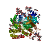
- PDB-7kvy: Crystal Structure of Acetyl-CoA synthetase in complex with adenos... -
+ Open data

- Basic information
| Entry | Database: PDB / ID: 7kvy | ||||||
|---|---|---|---|---|---|---|---|
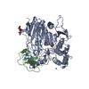
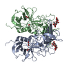
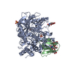
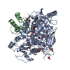
| Title | Crystal Structure of Acetyl-CoA synthetase in complex with adenosine-5'-ethylphosphate and Co-enzyme A from Coccidioides immitis RS | ||||||
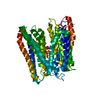
 Components Components | Acetyl-coenzyme A synthetase | ||||||
 Keywords Keywords | LIGASE / SSGCID / Acetyl-coenzyme A synthetase / ethyl-AMP / CoA / Co-Enzyme A / Structural Genomics / Seattle Structural Genomics Center for Infectious Disease | ||||||
| Function / homology |  Function and homology information Function and homology informationacetate-CoA ligase / acetyl-CoA synthetase activity / : / AMP binding / ATP binding / cytosol Similarity search - Function | ||||||
| Biological species |  Coccidioides immitis (fungus) Coccidioides immitis (fungus) | ||||||
| Method |  X-RAY DIFFRACTION / SYNCHROTRON / MOLECULAR REPLACEMENT / Resolution: 1.9 Å X-RAY DIFFRACTION / SYNCHROTRON / MOLECULAR REPLACEMENT / Resolution: 1.9 Å | ||||||
 Authors Authors | Seattle Structural Genomics Center for Infectious Disease (SSGCID) | ||||||
 Citation Citation | Journal: to be published Title: Crystal Structure of Acetyl-CoA synthetase in complex with adenosine-5'-ethylphosphate and Co-enzyme A from Coccidioides immitis RS Authors: Fox III, D. / Abendroth, J. / DeBouver, N.D. / Esan, T.E. / Hagen, T.J. / Krysan, D.J. / Lorimer, D.D. / Horanyi, P.S. / Edwards, T.E. | ||||||
| History |
|
- Structure visualization
| Structure viewer | Molecule: MolmilJmol/JSmol |
|---|
- Downloads & links
-Download
| PDBx/mmCIF format | 7kvy.cif.gz | 274.1 KB | Display | PDBx/mmCIF format |
|---|---|---|---|---|
| PDB format | pdb7kvy.ent.gz | 218.4 KB | Display | PDB format |
| PDBx/mmJSON format | 7kvy.json.gz | Tree view | PDBx/mmJSON format | |
| Others |  Other downloads Other downloads |
-Validation report
| Arichive directory | https://data.pdbj.org/pub/pdb/validation_reports/kv/7kvyftp://data.pdbj.org/pub/pdb/validation_reports/kv/7kvy | HTTPS FTP |
|---|
-Related structure data
| Related structure data | 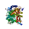 7kcpS S: Starting model for refinement |
|---|---|
| Similar structure data |


-Links
 PDBj
PDBj


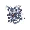
- Assembly
| Deposited unit | 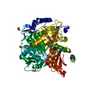
| ||||||||
|---|---|---|---|---|---|---|---|---|---|
| 1 |
| ||||||||
| Unit cell |
|
-Components
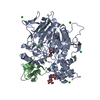
| #1: Protein | Mass: 78435.828 Da / Num. of mol.: 1 / Fragment: CoimA.00629.a.FS11 Source method: isolated from a genetically manipulated source Source: (gene. exp.) Coccidioides immitis (strain RS) (fungus)Strain: RS / Gene: CIMG_01510 / Plasmid: CoimA.00629.a.FS11 / Production host:  | ||||
|---|---|---|---|---|---|
| #2: Chemical | ChemComp-COA /   Mass: 767.534 Da / Num. of mol.: 1 / Source method: obtained synthetically / Formula: C21H36N7O16P3S Mass: 767.534 Da / Num. of mol.: 1 / Source method: obtained synthetically / Formula: C21H36N7O16P3S | ||||
| #3: Chemical | ChemComp-WTA / 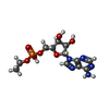  Mass: 375.274 Da / Num. of mol.: 1 / Source method: obtained synthetically / Formula: C12H18N5O7P Mass: 375.274 Da / Num. of mol.: 1 / Source method: obtained synthetically / Formula: C12H18N5O7P | ||||
| #4: Chemical | ChemComp-EDO /   Mass: 62.068 Da / Num. of mol.: 9 / Source method: obtained synthetically / Formula: C2H6O2 Mass: 62.068 Da / Num. of mol.: 9 / Source method: obtained synthetically / Formula: C2H6O2#5: Water | ChemComp-HOH / |  Mass: 18.015 Da / Num. of mol.: 344 / Source method: isolated from a natural source / Formula: H2O Mass: 18.015 Da / Num. of mol.: 344 / Source method: isolated from a natural source / Formula: H2OHas ligand of interest | Y | |
-Experimental details
-Experiment
| Experiment | Method: X-RAY DIFFRACTION / Number of used crystals: 1 |
|---|
- Sample preparation
| Crystal | Density Matthews: 2.45 Å3/Da / Density % sol: 49.8 % / Mosaicity: 0.18 ° |
|---|---|
| Crystal grow | Temperature: 287 K / Method: vapor diffusion, sitting drop / pH: 7 Details: Optimization conditions around RigakuReagents JCSG H3: 18.32@ PEG3,350, 0.2M Lithium acetate; CoimA.00629.a.FS11.PD00401 at 10 mg/mL + 1mM TCEP (added first) + 1mM adenosine-5'- ...Details: Optimization conditions around RigakuReagents JCSG H3: 18.32@ PEG3,350, 0.2M Lithium acetate; CoimA.00629.a.FS11.PD00401 at 10 mg/mL + 1mM TCEP (added first) + 1mM adenosine-5'-ethylphosphate + 1mM Co-EnzymeA; Cryo: 20% ethylene glycol; tray 317944f5, puck dzd7-5. |
-Data collection
| Diffraction | Mean temperature: 100 K / Serial crystal experiment: N |
|---|---|
| Diffraction source | Source: SYNCHROTRON / Site: APS  / Beamline: 21-ID-F / Wavelength: 0.97872 Å / Beamline: 21-ID-F / Wavelength: 0.97872 Å |
| Detector | Type: RAYONIX MX-300 / Detector: CCD / Date: Sep 24, 2020 / Details: Beryllium Lenses |
| Radiation | Monochromator: Diamond [111] / Protocol: SINGLE WAVELENGTH / Monochromatic (M) / Laue (L): M / Scattering type: x-ray |
| Radiation wavelength | Wavelength: 0.97872 Å / Relative weight: 1 |
| Reflection | Resolution: 1.9→50 Å / Num. obs: 59143 / % possible obs: 100 % / Observed criterion σ(I): -3 / Redundancy: 5.7 % / Biso Wilson estimate: 33.21 Å2 / Rmerge(I) obs: 0.053 / Net I/σ(I): 18.29 |
| Reflection shell | Resolution: 1.9→1.95 Å / Rmerge(I) obs: 0.86 / Mean I/σ(I) obs: 2.09 / % possible all: 100 |
- Processing
| Software |
| ||||||||||||||||||||||||||||||||||||||||||||||||||||||||||||||||||||||||||||||||||||||||||||||||||||||||||||||||||||||||||||||||||||||||||||||||||||||||||||||||||||||||||||||||||||||||||||||||||||||||
|---|---|---|---|---|---|---|---|---|---|---|---|---|---|---|---|---|---|---|---|---|---|---|---|---|---|---|---|---|---|---|---|---|---|---|---|---|---|---|---|---|---|---|---|---|---|---|---|---|---|---|---|---|---|---|---|---|---|---|---|---|---|---|---|---|---|---|---|---|---|---|---|---|---|---|---|---|---|---|---|---|---|---|---|---|---|---|---|---|---|---|---|---|---|---|---|---|---|---|---|---|---|---|---|---|---|---|---|---|---|---|---|---|---|---|---|---|---|---|---|---|---|---|---|---|---|---|---|---|---|---|---|---|---|---|---|---|---|---|---|---|---|---|---|---|---|---|---|---|---|---|---|---|---|---|---|---|---|---|---|---|---|---|---|---|---|---|---|---|---|---|---|---|---|---|---|---|---|---|---|---|---|---|---|---|---|---|---|---|---|---|---|---|---|---|---|---|---|---|---|---|---|
| Refinement | Method to determine structure: MOLECULAR REPLACEMENT Starting model: 7KCP Resolution: 1.9→46.31 Å / SU ML: 0.2 / Cross valid method: FREE R-VALUE / σ(F): 1.35 / Phase error: 20.35 / Stereochemistry target values: ML
| ||||||||||||||||||||||||||||||||||||||||||||||||||||||||||||||||||||||||||||||||||||||||||||||||||||||||||||||||||||||||||||||||||||||||||||||||||||||||||||||||||||||||||||||||||||||||||||||||||||||||
| Solvent computation | Shrinkage radii: 0.9 Å / VDW probe radii: 1.11 Å / Solvent model: FLAT BULK SOLVENT MODEL | ||||||||||||||||||||||||||||||||||||||||||||||||||||||||||||||||||||||||||||||||||||||||||||||||||||||||||||||||||||||||||||||||||||||||||||||||||||||||||||||||||||||||||||||||||||||||||||||||||||||||
| Displacement parameters | Biso mean: 47.05 Å2 | ||||||||||||||||||||||||||||||||||||||||||||||||||||||||||||||||||||||||||||||||||||||||||||||||||||||||||||||||||||||||||||||||||||||||||||||||||||||||||||||||||||||||||||||||||||||||||||||||||||||||
| Refinement step | Cycle: LAST / Resolution: 1.9→46.31 Å
| ||||||||||||||||||||||||||||||||||||||||||||||||||||||||||||||||||||||||||||||||||||||||||||||||||||||||||||||||||||||||||||||||||||||||||||||||||||||||||||||||||||||||||||||||||||||||||||||||||||||||
| Refine LS restraints |
| ||||||||||||||||||||||||||||||||||||||||||||||||||||||||||||||||||||||||||||||||||||||||||||||||||||||||||||||||||||||||||||||||||||||||||||||||||||||||||||||||||||||||||||||||||||||||||||||||||||||||
| LS refinement shell |
| ||||||||||||||||||||||||||||||||||||||||||||||||||||||||||||||||||||||||||||||||||||||||||||||||||||||||||||||||||||||||||||||||||||||||||||||||||||||||||||||||||||||||||||||||||||||||||||||||||||||||
| Refinement TLS params. | Method: refined / Refine-ID: X-RAY DIFFRACTION
| ||||||||||||||||||||||||||||||||||||||||||||||||||||||||||||||||||||||||||||||||||||||||||||||||||||||||||||||||||||||||||||||||||||||||||||||||||||||||||||||||||||||||||||||||||||||||||||||||||||||||
| Refinement TLS group |
|
