 Movie
Movie Controller

[English] 日本語
 Yorodumi
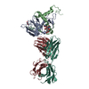
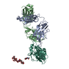
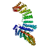
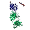
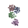
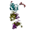
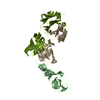
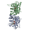
- PDB-7kq7: Crystal structure of IL21R in complex with an antibody Fab fragment -
+ Open data

- Basic information
| Entry | Database: PDB / ID: 7kq7 | ||||||
|---|---|---|---|---|---|---|---|
| Title | Crystal structure of IL21R in complex with an antibody Fab fragment | ||||||
 Components Components |
| ||||||
 Keywords Keywords | PROTEIN BINDING/Immune System / cytokine receptor / PROTEIN BINDING / PROTEIN BINDING-Immune System complex | ||||||
| Function / homology |  Function and homology information Function and homology informationinterleukin-21 receptor activity / Interleukin-21 signaling / natural killer cell activation / cytokine receptor activity / cytokine-mediated signaling pathway / transmembrane signaling receptor activity / external side of plasma membrane / membrane / plasma membrane Similarity search - Function | ||||||
| Biological species |   Homo sapiens (human) Homo sapiens (human) | ||||||
| Method |  X-RAY DIFFRACTION / SYNCHROTRON / MOLECULAR REPLACEMENT / Resolution: 2.203 Å X-RAY DIFFRACTION / SYNCHROTRON / MOLECULAR REPLACEMENT / Resolution: 2.203 Å | ||||||
 Authors Authors | Mosyak, L. / Svenson, K. | ||||||
 Citation Citation | Journal: Mabs / Year: 2021 Title: Combining random mutagenesis, structure-guided design and next-generation sequencing to mitigate polyreactivity of an anti-IL-21R antibody. Authors: Campbell, S.M. / DeBartolo, J. / Apgar, J.R. / Mosyak, L. / McManus, V. / Beyer, S. / Bennett, E.M. / Lambert, M. / Cunningham, O. | ||||||
| History |
|
- Structure visualization
| Structure viewer | Molecule: MolmilJmol/JSmol |
|---|
- Downloads & links
-Download
| PDBx/mmCIF format | 7kq7.cif.gz | 141.8 KB | Display | PDBx/mmCIF format |
|---|---|---|---|---|
| PDB format | pdb7kq7.ent.gz | 110.1 KB | Display | PDB format |
| PDBx/mmJSON format | 7kq7.json.gz | Tree view | PDBx/mmJSON format | |
| Others |  Other downloads Other downloads |
-Validation report
| Arichive directory | https://data.pdbj.org/pub/pdb/validation_reports/kq/7kq7ftp://data.pdbj.org/pub/pdb/validation_reports/kq/7kq7 | HTTPS FTP |
|---|
-Related structure data
| Similar structure data |
|---|


-Links
 PDBj
PDBj


- Assembly
| Deposited unit | 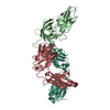
| ||||||||
|---|---|---|---|---|---|---|---|---|---|
| 1 |
| ||||||||
| Unit cell |
|
-Components
| #1: Antibody | Mass: 23848.814 Da / Num. of mol.: 1 Source method: isolated from a genetically manipulated source Source: (gene. exp.)  Cricetulus griseus (Chinese hamster) Cricetulus griseus (Chinese hamster) |
|---|---|
| #2: Antibody | Mass: 23777.381 Da / Num. of mol.: 1 Source method: isolated from a genetically manipulated source Source: (gene. exp.) Cricetulus griseus (Chinese hamster) |
| #3: Protein | Mass: 24636.406 Da / Num. of mol.: 1 Source method: isolated from a genetically manipulated source Source: (gene. exp.) Homo sapiens (human) / Gene: IL21R, NILR, UNQ3121/PRO10273 / Production host: Cricetulus griseus (Chinese hamster) / References: UniProt: Q9HBE5 |
| #4: Water | ChemComp-HOH /  Mass: 18.015 Da / Num. of mol.: 149 / Source method: isolated from a natural source / Formula: H2O Mass: 18.015 Da / Num. of mol.: 149 / Source method: isolated from a natural source / Formula: H2O |
| Has protein modification | Y |
-Experimental details
-Experiment
| Experiment | Method: X-RAY DIFFRACTION / Number of used crystals: 1 |
|---|
- Sample preparation
| Crystal | Density Matthews: 3.65 Å3/Da / Density % sol: 66.31 % |
|---|---|
| Crystal grow | Temperature: 300 K / Method: vapor diffusion, hanging drop / pH: 6.5 / Details: 10% PEG MME 5000 and 0.1M MES, pH 6.5 |
-Data collection
| Diffraction | Mean temperature: 230 K / Serial crystal experiment: N |
|---|---|
| Diffraction source | Source: SYNCHROTRON / Site: APS  / Beamline: 17-ID / Wavelength: 1 Å / Beamline: 17-ID / Wavelength: 1 Å |
| Detector | Type: DECTRIS PILATUS 300K / Detector: PIXEL / Date: May 5, 2010 |
| Radiation | Protocol: SINGLE WAVELENGTH / Monochromatic (M) / Laue (L): M / Scattering type: x-ray |
| Radiation wavelength | Wavelength: 1 Å / Relative weight: 1 |
| Reflection | Resolution: 2.2→50 Å / Num. obs: 47436 / % possible obs: 87.8 % / Redundancy: 4.6 % / Rmerge(I) obs: 0.1 / Net I/σ(I): 18.8 |
| Reflection shell | Resolution: 2.2→2.24 Å / Rmerge(I) obs: 0.58 / Num. unique obs: 1437 |
- Processing
| Software |
| ||||||||||||||||||||||||||||||||||||||||||||||||||||||||||||||||||
|---|---|---|---|---|---|---|---|---|---|---|---|---|---|---|---|---|---|---|---|---|---|---|---|---|---|---|---|---|---|---|---|---|---|---|---|---|---|---|---|---|---|---|---|---|---|---|---|---|---|---|---|---|---|---|---|---|---|---|---|---|---|---|---|---|---|---|---|
| Refinement | Method to determine structure: MOLECULAR REPLACEMENT Starting model: in house structure Resolution: 2.203→42.216 Å / SU ML: 0.28 / Cross valid method: THROUGHOUT / σ(F): 1.34 / Phase error: 25.93 / Stereochemistry target values: ML
| ||||||||||||||||||||||||||||||||||||||||||||||||||||||||||||||||||
| Solvent computation | Shrinkage radii: 0.9 Å / VDW probe radii: 1.11 Å / Solvent model: FLAT BULK SOLVENT MODEL / Bsol: 45.491 Å2 / ksol: 0.336 e/Å3 | ||||||||||||||||||||||||||||||||||||||||||||||||||||||||||||||||||
| Displacement parameters | Biso max: 147.77 Å2 / Biso mean: 67.69 Å2 / Biso min: 21.13 Å2
| ||||||||||||||||||||||||||||||||||||||||||||||||||||||||||||||||||
| Refinement step | Cycle: final / Resolution: 2.203→42.216 Å
| ||||||||||||||||||||||||||||||||||||||||||||||||||||||||||||||||||
| Refine LS restraints |
| ||||||||||||||||||||||||||||||||||||||||||||||||||||||||||||||||||
| LS refinement shell | Refine-ID: X-RAY DIFFRACTION / Rfactor Rfree error: 0
|
