 Movie
Movie Controller

[English] 日本語
 Yorodumi
- PDB-7kio: Crystal structure of inositol polyphosphate 1-phosphatase (INPP1)... -
+ Open data

- Basic information
| Entry | Database: PDB / ID: 7kio | ||||||||||||
|---|---|---|---|---|---|---|---|---|---|---|---|---|---|
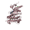
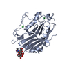
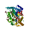
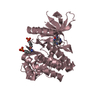
| Title | Crystal structure of inositol polyphosphate 1-phosphatase (INPP1) D54A mutant | ||||||||||||
 Components Components | Inositol polyphosphate 1-phosphatase | ||||||||||||
 Keywords Keywords | HYDROLASE | ||||||||||||
| Function / homology |  Function and homology information Function and homology informationinositol-1,3,4-trisphosphate 1-phosphatase activity / Synthesis of IP2, IP, and Ins in the cytosol / inositol-1,4-bisphosphate 1-phosphatase / inositol-1,4-bisphosphate 1-phosphatase activity / phosphatidylinositol phosphate biosynthetic process / metal ion binding Similarity search - Function | ||||||||||||
| Biological species |  | ||||||||||||
| Method |  X-RAY DIFFRACTION / MOLECULAR REPLACEMENT / Resolution: 2.4 Å X-RAY DIFFRACTION / MOLECULAR REPLACEMENT / Resolution: 2.4 Å | ||||||||||||
 Authors Authors | Xiong, J.-P. / Dollins, D.E. / Ren, Y. / York, J.D. | ||||||||||||
| Funding support |  United States, 3items United States, 3items
| ||||||||||||
 Citation Citation | Journal: J.Biol.Chem. / Year: 2020 Title: A structural basis for lithium and substrate binding of an inositide phosphatase. Authors: Dollins, D.E. / Xiong, J.P. / Endo-Streeter, S. / Anderson, D.E. / Bansal, V.S. / Ponder, J.W. / Ren, Y. / York, J.D. | ||||||||||||
| History |
|
- Structure visualization
| Structure viewer | Molecule: MolmilJmol/JSmol |
|---|
- Downloads & links
-Download
| PDBx/mmCIF format | 7kio.cif.gz | 95.7 KB | Display | PDBx/mmCIF format |
|---|---|---|---|---|
| PDB format | pdb7kio.ent.gz | 57.3 KB | Display | PDB format |
| PDBx/mmJSON format | 7kio.json.gz | Tree view | PDBx/mmJSON format | |
| Others |  Other downloads Other downloads |
-Validation report
| Arichive directory | https://data.pdbj.org/pub/pdb/validation_reports/ki/7kioftp://data.pdbj.org/pub/pdb/validation_reports/ki/7kio | HTTPS FTP |
|---|
-Related structure data
| Related structure data |  6wroC 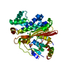 6wrrC  6wryC  6x25C 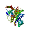 7kirC  1inpS S: Starting model for refinement C: citing same article ( |
|---|---|
| Similar structure data |


-Links
 PDBj
PDBj- Assembly
| Deposited unit | 
| ||||||||||||
|---|---|---|---|---|---|---|---|---|---|---|---|---|---|
| 1 |
| ||||||||||||
| Unit cell |
|
-Components
| #1: Protein | Mass: 43932.922 Da / Num. of mol.: 1 / Mutation: D54A Source method: isolated from a genetically manipulated source Source: (gene. exp.)   Spodoptera frugiperda (fall armyworm) Spodoptera frugiperda (fall armyworm)References: UniProt: P21327, inositol-1,4-bisphosphate 1-phosphatase | ||||
|---|---|---|---|---|---|
| #2: Chemical | ChemComp-CA /   Mass: 40.078 Da / Num. of mol.: 1 / Source method: obtained synthetically / Formula: Ca Mass: 40.078 Da / Num. of mol.: 1 / Source method: obtained synthetically / Formula: Ca | ||||
| #3: Chemical |   Mass: 96.063 Da / Num. of mol.: 2 / Source method: obtained synthetically / Formula: SO4 Mass: 96.063 Da / Num. of mol.: 2 / Source method: obtained synthetically / Formula: SO4#4: Water | ChemComp-HOH / |  Mass: 18.015 Da / Num. of mol.: 47 / Source method: isolated from a natural source / Formula: H2O Mass: 18.015 Da / Num. of mol.: 47 / Source method: isolated from a natural source / Formula: H2OHas ligand of interest | N | |
-Experimental details
-Experiment
| Experiment | Method: X-RAY DIFFRACTION / Number of used crystals: 1 |
|---|
- Sample preparation
| Crystal | Density Matthews: 2.16 Å3/Da / Density % sol: 42.94 % |
|---|---|
| Crystal grow | Temperature: 293 K / Method: vapor diffusion, hanging drop Details: 0.1 M Sodium Citrate pH 4.3, 16-18% PEG6000, 5 mM CaCl2 |
-Data collection
| Diffraction | Mean temperature: 277 K / Serial crystal experiment: N |
|---|---|
| Diffraction source | Source: ROTATING ANODE / Type: RIGAKU R-AXIS II / Wavelength: 1.5418 Å |
| Detector | Type: RIGAKU RAXIS IIC / Detector: IMAGE PLATE / Date: Jan 1, 1998 |
| Radiation | Protocol: SINGLE WAVELENGTH / Monochromatic (M) / Laue (L): M / Scattering type: x-ray |
| Radiation wavelength | Wavelength: 1.5418 Å / Relative weight: 1 |
| Reflection | Resolution: 2.4→28.93 Å / Num. obs: 14352 / % possible obs: 98.6 % / Redundancy: 3.2 % / Biso Wilson estimate: 32.6 Å2 / Rmerge(I) obs: 0.09 / Net I/σ(I): 15.4 |
| Reflection shell | Resolution: 2.4→2.49 Å / Rmerge(I) obs: 0.401 / Num. unique obs: 1311 |
- Processing
| Software |
| |||||||||||||||||||||||||||||||||||||||||||||||||
|---|---|---|---|---|---|---|---|---|---|---|---|---|---|---|---|---|---|---|---|---|---|---|---|---|---|---|---|---|---|---|---|---|---|---|---|---|---|---|---|---|---|---|---|---|---|---|---|---|---|---|
| Refinement | Method to determine structure: MOLECULAR REPLACEMENT Starting model: 1INP Resolution: 2.4→28.93 Å / SU ML: 0.206 / Cross valid method: FREE R-VALUE / σ(F): 0 / Phase error: 25.7232 Stereochemistry target values: GeoStd + Monomer Library + CDL v1.2
| |||||||||||||||||||||||||||||||||||||||||||||||||
| Solvent computation | Shrinkage radii: 0.9 Å / VDW probe radii: 1.11 Å / Solvent model: FLAT BULK SOLVENT MODEL | |||||||||||||||||||||||||||||||||||||||||||||||||
| Displacement parameters | Biso mean: 38.85 Å2 | |||||||||||||||||||||||||||||||||||||||||||||||||
| Refinement step | Cycle: LAST / Resolution: 2.4→28.93 Å
| |||||||||||||||||||||||||||||||||||||||||||||||||
| Refine LS restraints |
| |||||||||||||||||||||||||||||||||||||||||||||||||
| LS refinement shell |
|
