 Movie
Movie Controller

[English] 日本語
 Yorodumi
- PDB-6wrr: CRYSTAL STRUCTURE OF INOSITOL POLYPHOSPHATE 1-PHOSPHATASE INPP1 I... -
+ Open data

- Basic information
| Entry | Database: PDB / ID: 6wrr | ||||||||||||
|---|---|---|---|---|---|---|---|---|---|---|---|---|---|
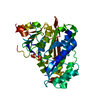
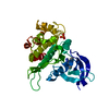
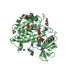
| Title | CRYSTAL STRUCTURE OF INOSITOL POLYPHOSPHATE 1-PHOSPHATASE INPP1 IN COMPLEX GADOLINIUM AND LITHIUM AT 2.5 ANGSTROM RESOLUTION | ||||||||||||
 Components Components | Inositol polyphosphate 1-phosphatase | ||||||||||||
 Keywords Keywords | SIGNALING PROTEIN / inositol phosphate / neurological disease / Bipolar disorder / manic depressive illness / phosphatase | ||||||||||||
| Function / homology |  Function and homology information Function and homology informationinositol-1,3,4-trisphosphate 1-phosphatase activity / Synthesis of IP2, IP, and Ins in the cytosol / inositol-1,4-bisphosphate 1-phosphatase / inositol-1,4-bisphosphate 1-phosphatase activity / phosphatidylinositol phosphate biosynthetic process / metal ion binding Similarity search - Function | ||||||||||||
| Biological species |  | ||||||||||||
| Method |  X-RAY DIFFRACTION / FOURIER SYNTHESIS / Resolution: 2.5 Å X-RAY DIFFRACTION / FOURIER SYNTHESIS / Resolution: 2.5 Å | ||||||||||||
 Authors Authors | Dollins, D.R. / Endo-Streeter, S. / York, J.D. | ||||||||||||
| Funding support |  United States, 3items United States, 3items
| ||||||||||||
 Citation Citation | Journal: J.Biol.Chem. / Year: 2020 Title: A structural basis for lithium and substrate binding of an inositide phosphatase. Authors: Dollins, D.E. / Xiong, J.P. / Endo-Streeter, S. / Anderson, D.E. / Bansal, V.S. / Ponder, J.W. / Ren, Y. / York, J.D. | ||||||||||||
| History |
|
- Structure visualization
| Structure viewer | Molecule: MolmilJmol/JSmol |
|---|
- Downloads & links
-Download
| PDBx/mmCIF format | 6wrr.cif.gz | 83.9 KB | Display | PDBx/mmCIF format |
|---|---|---|---|---|
| PDB format | pdb6wrr.ent.gz | 59.5 KB | Display | PDB format |
| PDBx/mmJSON format | 6wrr.json.gz | Tree view | PDBx/mmJSON format | |
| Others |  Other downloads Other downloads |
-Validation report
| Arichive directory | https://data.pdbj.org/pub/pdb/validation_reports/wr/6wrrftp://data.pdbj.org/pub/pdb/validation_reports/wr/6wrr | HTTPS FTP |
|---|
-Related structure data
| Related structure data |  6wroC  6wryC  6x25C 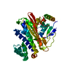 7kioC 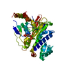 7kirC  1inpS S: Starting model for refinement C: citing same article ( |
|---|---|
| Similar structure data |


-Links
 PDBj
PDBj- Assembly
| Deposited unit | 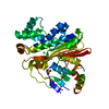
| ||||||||
|---|---|---|---|---|---|---|---|---|---|
| 1 |
| ||||||||
| Unit cell |
|
-Components
| #1: Protein | Mass: 43976.934 Da / Num. of mol.: 1 Source method: isolated from a genetically manipulated source Source: (gene. exp.)  Baculoviridae sp. (virus) Baculoviridae sp. (virus)References: UniProt: P21327, inositol-1,4-bisphosphate 1-phosphatase | ||||||
|---|---|---|---|---|---|---|---|
| #2: Chemical |   Mass: 157.250 Da / Num. of mol.: 2 / Source method: obtained synthetically / Formula: Gd / Feature type: SUBJECT OF INVESTIGATION Mass: 157.250 Da / Num. of mol.: 2 / Source method: obtained synthetically / Formula: Gd / Feature type: SUBJECT OF INVESTIGATION#3: Chemical |   Mass: 96.063 Da / Num. of mol.: 2 / Source method: obtained synthetically / Formula: SO4 Mass: 96.063 Da / Num. of mol.: 2 / Source method: obtained synthetically / Formula: SO4#4: Water | ChemComp-HOH / |  Mass: 18.015 Da / Num. of mol.: 85 / Source method: isolated from a natural source / Formula: H2O Mass: 18.015 Da / Num. of mol.: 85 / Source method: isolated from a natural source / Formula: H2OHas ligand of interest | Y | |
-Experimental details
-Experiment
| Experiment | Method: X-RAY DIFFRACTION / Number of used crystals: 1 |
|---|
- Sample preparation
| Crystal | Density Matthews: 2.17 Å3/Da / Density % sol: 43.39 % Description: Tetragonal shaped crystals amenable to diffraction studies were grown by the hanging drop vapor diffusion method on silanized glass covers slips at 20 C and grew with dimensions ...Description: Tetragonal shaped crystals amenable to diffraction studies were grown by the hanging drop vapor diffusion method on silanized glass covers slips at 20 C and grew with dimensions routinely exceeding 0.1 x 0.1 x 0.5mm. Gd3+, a competitive inhibitor of Mg2+, has a useful anomalous absorption edge at the CuK wavelength and therefore was used in the co-crystallization or soaking conditions. |
|---|---|
| Crystal grow | Temperature: 293 K / Method: vapor diffusion, hanging drop / pH: 6.3 Details: 13% PEG8000, 200mM Li2SO4 and 100mM Tris, pH 6.3. Upon reaching maximum size after ~5 days, a Gd3+ heavy atom derivative was prepared by adding 0.5uL of 17.5mM Gd2(SO4)3 in reservoir ...Details: 13% PEG8000, 200mM Li2SO4 and 100mM Tris, pH 6.3. Upon reaching maximum size after ~5 days, a Gd3+ heavy atom derivative was prepared by adding 0.5uL of 17.5mM Gd2(SO4)3 in reservoir solution to 3uL hanging drops (2.5mM final Gd3+) containing native crystals for 48 hours |
-Data collection
| Diffraction | Mean temperature: 296 K / Serial crystal experiment: N |
|---|---|
| Diffraction source | Source: ROTATING ANODE / Type: RIGAKU RU200 / Wavelength: 1.542 Å |
| Detector | Type: UCSD MARK II / Detector: AREA DETECTOR / Date: Jul 10, 1992 |
| Radiation | Monochromator: supper graphite / Protocol: SINGLE WAVELENGTH / Monochromatic (M) / Laue (L): M / Scattering type: x-ray |
| Radiation wavelength | Wavelength: 1.542 Å / Relative weight: 1 |
| Reflection | Resolution: 2.5→15.45 Å / Num. obs: 26197 / % possible obs: 96 % / Redundancy: 3.4 % / Rsym value: 0.051 / Net I/σ(I): 10.8 |
| Reflection shell | Resolution: 2.5→2.69 Å / Num. unique obs: 2250 / Rsym value: 0.172 / % possible all: 95.6 |
- Processing
| Software |
| ||||||||||||||||||||||||||||||||||||||||||||||||||||||||||||||||||||||||||||||||||||||||||||||||||||||||||||||||||||||||||||||||||||||||||||||||||||||||||||||||||||||||||||||||||||||
|---|---|---|---|---|---|---|---|---|---|---|---|---|---|---|---|---|---|---|---|---|---|---|---|---|---|---|---|---|---|---|---|---|---|---|---|---|---|---|---|---|---|---|---|---|---|---|---|---|---|---|---|---|---|---|---|---|---|---|---|---|---|---|---|---|---|---|---|---|---|---|---|---|---|---|---|---|---|---|---|---|---|---|---|---|---|---|---|---|---|---|---|---|---|---|---|---|---|---|---|---|---|---|---|---|---|---|---|---|---|---|---|---|---|---|---|---|---|---|---|---|---|---|---|---|---|---|---|---|---|---|---|---|---|---|---|---|---|---|---|---|---|---|---|---|---|---|---|---|---|---|---|---|---|---|---|---|---|---|---|---|---|---|---|---|---|---|---|---|---|---|---|---|---|---|---|---|---|---|---|---|---|---|---|
| Refinement | Method to determine structure: FOURIER SYNTHESIS Starting model: 1INP Resolution: 2.5→15.45 Å / Cor.coef. Fo:Fc: 0.95 / Cor.coef. Fo:Fc free: 0.935 / SU B: 13.952 / SU ML: 0.168 / Cross valid method: THROUGHOUT / ESU R: 0.616 / ESU R Free: 0.248 / Details: HYDROGENS HAVE BEEN ADDED IN THE RIDING POSITIONS
| ||||||||||||||||||||||||||||||||||||||||||||||||||||||||||||||||||||||||||||||||||||||||||||||||||||||||||||||||||||||||||||||||||||||||||||||||||||||||||||||||||||||||||||||||||||||
| Solvent computation | Ion probe radii: 0.8 Å / Shrinkage radii: 0.8 Å / VDW probe radii: 1.2 Å | ||||||||||||||||||||||||||||||||||||||||||||||||||||||||||||||||||||||||||||||||||||||||||||||||||||||||||||||||||||||||||||||||||||||||||||||||||||||||||||||||||||||||||||||||||||||
| Displacement parameters | Biso mean: 32.228 Å2
| ||||||||||||||||||||||||||||||||||||||||||||||||||||||||||||||||||||||||||||||||||||||||||||||||||||||||||||||||||||||||||||||||||||||||||||||||||||||||||||||||||||||||||||||||||||||
| Refinement step | Cycle: 1 / Resolution: 2.5→15.45 Å
| ||||||||||||||||||||||||||||||||||||||||||||||||||||||||||||||||||||||||||||||||||||||||||||||||||||||||||||||||||||||||||||||||||||||||||||||||||||||||||||||||||||||||||||||||||||||
| Refine LS restraints |
|