 Movie
Movie Controller

[English] 日本語
 Yorodumi
- PDB-1inp: CRYSTAL STRUCTURE OF INOSITOL POLYPHOSPHATE 1-PHOSPHATASE AT 2.3 ... -
+ Open data

- Basic information
| Entry | Database: PDB / ID: 1inp | ||||||
|---|---|---|---|---|---|---|---|
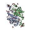
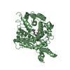
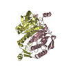
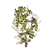
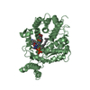
| Title | CRYSTAL STRUCTURE OF INOSITOL POLYPHOSPHATE 1-PHOSPHATASE AT 2.3 ANGSTROMS RESOLUTION | ||||||
 Components Components | INOSITOL POLYPHOSPHATE 1-PHOSPHATASE | ||||||
 Keywords Keywords | HYDROLASE(PHOSPHORIC MONOESTER) | ||||||
| Function / homology |  Function and homology information Function and homology informationinositol-1,3,4-trisphosphate 1-phosphatase activity / Synthesis of IP2, IP, and Ins in the cytosol / inositol-1,4-bisphosphate 1-phosphatase / inositol-1,4-bisphosphate 1-phosphatase activity / phosphatidylinositol phosphate biosynthetic process / metal ion binding Similarity search - Function | ||||||
| Biological species |  | ||||||
| Method |  X-RAY DIFFRACTION / Resolution: 2.3 Å X-RAY DIFFRACTION / Resolution: 2.3 Å | ||||||
 Authors Authors | York, J.D. / Ponder, J.W. / Chen, Z. / Mathews, F.S. / Majerus, P.W. | ||||||
 Citation Citation | Journal: Biochemistry / Year: 1994 Title: Crystal structure of inositol polyphosphate 1-phosphatase at 2.3-A resolution. Authors: York, J.D. / Ponder, J.W. / Chen, Z.W. / Mathews, F.S. / Majerus, P.W. #1: Journal: J.Mol.Biol. / Year: 1994Title: Crystallization and Initial X-Ray Crystallographic Characterization of Recombinant Bovine Inositol Polyphosphate 1-Phosphatase Produced in Spodoptera Frugiperda Cells Authors: York, J.D. / Chen, Z. / Ponder, J.W. / Chauhan, A.K. / Mathews, F.S. / Majerus, P.W. | ||||||
| History |
|
- Structure visualization
| Structure viewer | Molecule: MolmilJmol/JSmol |
|---|
- Downloads & links
-Download
| PDBx/mmCIF format | 1inp.cif.gz | 108 KB | Display | PDBx/mmCIF format |
|---|---|---|---|---|
| PDB format | pdb1inp.ent.gz | 83.4 KB | Display | PDB format |
| PDBx/mmJSON format | 1inp.json.gz | Tree view | PDBx/mmJSON format | |
| Others |  Other downloads Other downloads |
-Validation report
| Arichive directory | https://data.pdbj.org/pub/pdb/validation_reports/in/1inpftp://data.pdbj.org/pub/pdb/validation_reports/in/1inp | HTTPS FTP |
|---|
-Related structure data
| Similar structure data |
|---|


-Links
 PDBj
PDBj- Assembly
| Deposited unit | 
| ||||||||
|---|---|---|---|---|---|---|---|---|---|
| 1 |
| ||||||||
| Unit cell |
| ||||||||
| Atom site foot note | 1: CIS PROLINE - PRO 134 2: LEU 234 - PRO 235 OMEGA = 136.45 PEPTIDE BOND DEVIATES SIGNIFICANTLY FROM TRANS CONFORMATION |
-Components
| #1: Protein | Mass: 43976.934 Da / Num. of mol.: 1 Source method: isolated from a genetically manipulated source Source: (gene. exp.)   Spodoptera frugiperda (fall armyworm) Spodoptera frugiperda (fall armyworm)References: UniProt: P21327, inositol-1,4-bisphosphate 1-phosphatase | ||||
|---|---|---|---|---|---|
| #2: Chemical |   Mass: 24.305 Da / Num. of mol.: 2 / Source method: obtained synthetically / Formula: Mg Mass: 24.305 Da / Num. of mol.: 2 / Source method: obtained synthetically / Formula: Mg#3: Water | ChemComp-HOH / |  Mass: 18.015 Da / Num. of mol.: 70 / Source method: isolated from a natural source / Formula: H2O Mass: 18.015 Da / Num. of mol.: 70 / Source method: isolated from a natural source / Formula: H2OCompound details | THE STRUCTURE OF 1-PTASE IS SIMILAR TO TWO OTHER MAGNESIUM DEPENDENT/LITHIUM SENSITIVE PHOSPHATASES: ...THE STRUCTURE OF 1-PTASE IS SIMILAR TO TWO OTHER MAGNESIUM DEPENDENT/LITHIUM SENSITIVE PHOSPHATAS | |
-Experimental details
-Experiment
| Experiment | Method: X-RAY DIFFRACTION |
|---|
- Sample preparation
| Crystal | Density Matthews: 2.17 Å3/Da / Density % sol: 43.37 % | ||||||||||||||||||||||||||||||||||||||||||||||||||||||||||||
|---|---|---|---|---|---|---|---|---|---|---|---|---|---|---|---|---|---|---|---|---|---|---|---|---|---|---|---|---|---|---|---|---|---|---|---|---|---|---|---|---|---|---|---|---|---|---|---|---|---|---|---|---|---|---|---|---|---|---|---|---|---|
| Crystal grow | *PLUS Temperature: 20 ℃ / Method: vapor diffusion, hanging drop | ||||||||||||||||||||||||||||||||||||||||||||||||||||||||||||
| Components of the solutions | *PLUS
|
-Data collection
| Radiation | Scattering type: x-ray |
|---|---|
| Radiation wavelength | Relative weight: 1 |
| Reflection | Num. obs: 17652 / Observed criterion σ(I): 2 |
| Reflection | *PLUS Highest resolution: 2.3 Å / % possible obs: 99.3 % / Num. measured all: 63687 / Rmerge F obs: 0.049 |
- Processing
| Software |
| ||||||||||||||||||||||||||||||||||||||||||||||||||||||||||||
|---|---|---|---|---|---|---|---|---|---|---|---|---|---|---|---|---|---|---|---|---|---|---|---|---|---|---|---|---|---|---|---|---|---|---|---|---|---|---|---|---|---|---|---|---|---|---|---|---|---|---|---|---|---|---|---|---|---|---|---|---|---|
| Refinement | Resolution: 2.3→8 Å / σ(F): 2
| ||||||||||||||||||||||||||||||||||||||||||||||||||||||||||||
| Refinement step | Cycle: LAST / Resolution: 2.3→8 Å
| ||||||||||||||||||||||||||||||||||||||||||||||||||||||||||||
| Refine LS restraints |
| ||||||||||||||||||||||||||||||||||||||||||||||||||||||||||||
| Software | *PLUS Name: X-PLOR / Classification: refinement | ||||||||||||||||||||||||||||||||||||||||||||||||||||||||||||
| Refinement | *PLUS Rfactor obs: 0.198 / Rfactor Rwork: 0.198 | ||||||||||||||||||||||||||||||||||||||||||||||||||||||||||||
| Solvent computation | *PLUS | ||||||||||||||||||||||||||||||||||||||||||||||||||||||||||||
| Displacement parameters | *PLUS Biso mean: 36.28 Å2 | ||||||||||||||||||||||||||||||||||||||||||||||||||||||||||||
| Refine LS restraints | *PLUS
|
