Protocol: SINGLE WAVELENGTH / Monochromatic (M) / Laue (L): M / Scattering type: x-ray
Radiation wavelength
Wavelength: 0.9 Å / Relative weight: 1
Reflection
Resolution: 2.5→40 Å / Num. obs: 28181 / % possible obs: 84.8 % / Redundancy: 4.4 % / Net I/σ(I): 14.2
-
Processing
Software
Name
Version
Classification
REFMAC
5.7.0029
refinement
HKL-2000
datareduction
HKL-2000
datascaling
MOLREP
phasing
Refinement
Resolution: 2.51→36.33 Å / Cor.coef. Fo:Fc: 0.934 / Cor.coef. Fo:Fc free: 0.864 / SU B: 9.645 / SU ML: 0.225 / Cross valid method: THROUGHOUT / ESU R Free: 0.093 / Details: HYDROGENS HAVE BEEN ADDED IN THE RIDING POSITIONS
Rfactor
Num. reflection
% reflection
Selection details
Rfree
0.30048
1433
5.1 %
RANDOM
Rwork
0.21028
-
-
-
obs
0.21476
26722
84.15 %
-
Solvent computation
Ion probe radii: 0.8 Å / Shrinkage radii: 0.8 Å / VDW probe radii: 1.4 Å
 Movie
Movie Controller


 Open data
 Basic information
 Components
 Keywords
 Function and homology information
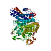
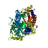
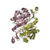
 Aeropyrum pernix (archaea)
 X-RAY DIFFRACTION /
  Authors
 Japan, 2items
  Citation
 Structure visualization
 Downloads & links
 Other downloads


 PDBj
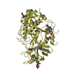
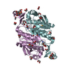
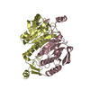
PDBj Assembly
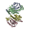


 Mass: 18.015 Da / Num. of mol.: 4 / Source method: isolated from a natural source / Formula: H2O
 Sample preparation
 Processing