 Movie
Movie Controller


+ Open data

- Basic information
| Entry | Database: PDB / ID: 7khm | ||||||
|---|---|---|---|---|---|---|---|
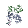
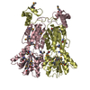
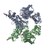
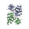
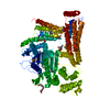
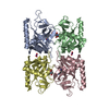
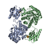
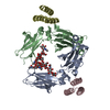
| Title | Crystal structure of hDHHS20 bound to palmitoyl CoA | ||||||
 Components Components | Isoform 4 of Palmitoyltransferase ZDHHC20 | ||||||
 Keywords Keywords | MEMBRANE PROTEIN / TRANSFERASE / DHHC / LIPID / ACYL / PALMITOYLTRANSFERASE | ||||||
| Function / homology |  Function and homology information Function and homology informationprotein-cysteine S-myristoyltransferase activity / protein-cysteine S-stearoyltransferase activity / peptidyl-L-cysteine S-palmitoylation / protein palmitoylation / protein S-acyltransferase / protein-cysteine S-palmitoyltransferase activity / palmitoyltransferase activity / synaptic vesicle maturation / host-mediated activation of viral process / protein targeting to membrane ...protein-cysteine S-myristoyltransferase activity / protein-cysteine S-stearoyltransferase activity / peptidyl-L-cysteine S-palmitoylation / protein palmitoylation / protein S-acyltransferase / protein-cysteine S-palmitoyltransferase activity / palmitoyltransferase activity / synaptic vesicle maturation / host-mediated activation of viral process / protein targeting to membrane / endoplasmic reticulum-Golgi intermediate compartment membrane / Transferases; Acyltransferases; Transferring groups other than aminoacyl groups / Maturation of spike protein / Golgi membrane / endoplasmic reticulum membrane / perinuclear region of cytoplasm / Golgi apparatus / endoplasmic reticulum / zinc ion binding / membrane / plasma membrane Similarity search - Function | ||||||
| Biological species |  Homo sapiens (human) Homo sapiens (human) | ||||||
| Method |  X-RAY DIFFRACTION / SYNCHROTRON / MOLECULAR REPLACEMENT / molecular replacement / Resolution: 2.88 Å X-RAY DIFFRACTION / SYNCHROTRON / MOLECULAR REPLACEMENT / molecular replacement / Resolution: 2.88 Å | ||||||
 Authors Authors | Lee, C.-J. / Banerjee, A. | ||||||
 Citation Citation | Journal: Proc.Natl.Acad.Sci.USA / Year: 2022 Title: Bivalent recognition of fatty acyl-CoA by a human integral membrane palmitoyltransferase. Authors: Lee, C.J. / Stix, R. / Rana, M.S. / Shikwana, F. / Murphy, R.E. / Ghirlando, R. / Faraldo-Gomez, J.D. / Banerjee, A. | ||||||
| History |
|
- Structure visualization
| Structure viewer | Molecule: MolmilJmol/JSmol |
|---|
- Downloads & links
-Download
| PDBx/mmCIF format | 7khm.cif.gz | 250.3 KB | Display | PDBx/mmCIF format |
|---|---|---|---|---|
| PDB format | pdb7khm.ent.gz | 199.2 KB | Display | PDB format |
| PDBx/mmJSON format | 7khm.json.gz | Tree view | PDBx/mmJSON format | |
| Others |  Other downloads Other downloads |
-Validation report
| Arichive directory | https://data.pdbj.org/pub/pdb/validation_reports/kh/7khmftp://data.pdbj.org/pub/pdb/validation_reports/kh/7khm | HTTPS FTP |
|---|
-Related structure data
| Related structure data |  6bmmS S: Starting model for refinement |
|---|---|
| Similar structure data |
-Links
 PDBj
PDBj- Assembly
| Deposited unit | 
| ||||||||
|---|---|---|---|---|---|---|---|---|---|
| 1 |
| ||||||||
| Unit cell |
|
-Components
| #1: Protein | Mass: 38494.324 Da / Num. of mol.: 2 / Mutation: C156S Source method: isolated from a genetically manipulated source Source: (gene. exp.) Homo sapiens (human) / Gene: ZDHHC20 / Production host:  Komagataella pastoris (fungus) Komagataella pastoris (fungus)References: UniProt: Q5W0Z9, protein S-acyltransferase , Transferases; Acyltransferases; Transferring groups other than aminoacyl groups #2: Chemical | ChemComp-ZN /   Mass: 65.409 Da / Num. of mol.: 4 / Source method: obtained synthetically / Formula: Zn Mass: 65.409 Da / Num. of mol.: 4 / Source method: obtained synthetically / Formula: Zn#3: Chemical |   Mass: 1005.943 Da / Num. of mol.: 2 / Source method: obtained synthetically / Formula: C37H66N7O17P3S / Feature type: SUBJECT OF INVESTIGATION Mass: 1005.943 Da / Num. of mol.: 2 / Source method: obtained synthetically / Formula: C37H66N7O17P3S / Feature type: SUBJECT OF INVESTIGATION#4: Chemical | ChemComp-PO4 / |   Mass: 94.971 Da / Num. of mol.: 1 / Source method: obtained synthetically / Formula: PO4 Mass: 94.971 Da / Num. of mol.: 1 / Source method: obtained synthetically / Formula: PO4#5: Water | ChemComp-HOH / |  Mass: 18.015 Da / Num. of mol.: 14 / Source method: isolated from a natural source / Formula: H2O Mass: 18.015 Da / Num. of mol.: 14 / Source method: isolated from a natural source / Formula: H2OHas ligand of interest | Y | |
|---|
-Experimental details
-Experiment
| Experiment | Method: X-RAY DIFFRACTION / Number of used crystals: 1 |
|---|
- Sample preparation
| Crystal | Density Matthews: 3.33 Å3/Da / Density % sol: 63.06 % |
|---|---|
| Crystal grow | Temperature: 293 K / Method: lipidic cubic phase Details: 50mM MES, pH 6.5, 50mM NaH2PO4, 30.3% PEG 300, 50mM DTT, 2.5% 2,5-hexanediol |
-Data collection
| Diffraction | Mean temperature: 100 K / Serial crystal experiment: N | ||||||||||||||||||||||||||||||
|---|---|---|---|---|---|---|---|---|---|---|---|---|---|---|---|---|---|---|---|---|---|---|---|---|---|---|---|---|---|---|---|
| Diffraction source | Source: SYNCHROTRON / Site: APS  / Beamline: 23-ID-D / Wavelength: 0.97918 Å / Beamline: 23-ID-D / Wavelength: 0.97918 Å | ||||||||||||||||||||||||||||||
| Detector | Type: DECTRIS EIGER X 16M / Detector: PIXEL / Date: Mar 17, 2019 | ||||||||||||||||||||||||||||||
| Radiation | Protocol: SINGLE WAVELENGTH / Monochromatic (M) / Laue (L): M / Scattering type: x-ray | ||||||||||||||||||||||||||||||
| Radiation wavelength | Wavelength: 0.97918 Å / Relative weight: 1 | ||||||||||||||||||||||||||||||
| Reflection | Resolution: 2.88→83.58 Å / Num. obs: 22858 / % possible obs: 99.7 % / Redundancy: 6.9 % / CC1/2: 0.999 / Rmerge(I) obs: 0.163 / Rpim(I) all: 0.067 / Rrim(I) all: 0.176 / Net I/σ(I): 7.6 / Num. measured all: 158295 | ||||||||||||||||||||||||||||||
| Reflection shell | Diffraction-ID: 1
|
-Phasing
| Phasing | Method: molecular replacement | |||||||||
|---|---|---|---|---|---|---|---|---|---|---|
| Phasing MR |
|
- Processing
| Software |
| |||||||||||||||||||||||||||||||||||||||||||||||||||||||||||||||||||||||||||||||||||||||||||||||||||||||||
|---|---|---|---|---|---|---|---|---|---|---|---|---|---|---|---|---|---|---|---|---|---|---|---|---|---|---|---|---|---|---|---|---|---|---|---|---|---|---|---|---|---|---|---|---|---|---|---|---|---|---|---|---|---|---|---|---|---|---|---|---|---|---|---|---|---|---|---|---|---|---|---|---|---|---|---|---|---|---|---|---|---|---|---|---|---|---|---|---|---|---|---|---|---|---|---|---|---|---|---|---|---|---|---|---|---|---|
| Refinement | Method to determine structure: MOLECULAR REPLACEMENT Starting model: 6BMM Resolution: 2.88→67.53 Å / SU ML: 0.56 / Cross valid method: THROUGHOUT / σ(F): 1.35 / Phase error: 44.85 / Stereochemistry target values: ML
| |||||||||||||||||||||||||||||||||||||||||||||||||||||||||||||||||||||||||||||||||||||||||||||||||||||||||
| Solvent computation | Shrinkage radii: 0.9 Å / VDW probe radii: 1.11 Å / Solvent model: FLAT BULK SOLVENT MODEL | |||||||||||||||||||||||||||||||||||||||||||||||||||||||||||||||||||||||||||||||||||||||||||||||||||||||||
| Displacement parameters | Biso max: 266.98 Å2 / Biso mean: 129.9206 Å2 / Biso min: 87.49 Å2 | |||||||||||||||||||||||||||||||||||||||||||||||||||||||||||||||||||||||||||||||||||||||||||||||||||||||||
| Refinement step | Cycle: final / Resolution: 2.88→67.53 Å
| |||||||||||||||||||||||||||||||||||||||||||||||||||||||||||||||||||||||||||||||||||||||||||||||||||||||||
| Refine LS restraints |
| |||||||||||||||||||||||||||||||||||||||||||||||||||||||||||||||||||||||||||||||||||||||||||||||||||||||||
| LS refinement shell | Refine-ID: X-RAY DIFFRACTION / Rfactor Rfree error: 0 / Total num. of bins used: 14
| |||||||||||||||||||||||||||||||||||||||||||||||||||||||||||||||||||||||||||||||||||||||||||||||||||||||||
| Refinement TLS params. | Method: refined / Origin x: 6.6658 Å / Origin y: 0.2702 Å / Origin z: 19.7347 Å
| |||||||||||||||||||||||||||||||||||||||||||||||||||||||||||||||||||||||||||||||||||||||||||||||||||||||||
| Refinement TLS group |
|
