 Movie
Movie Controller


+ Open data

- Basic information
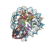
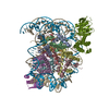
| Entry | Database: PDB / ID: 7kdp | ||||||
|---|---|---|---|---|---|---|---|
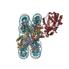
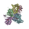
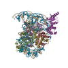
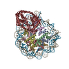
| Title | HCMV prefusion gB in complex with fusion inhibitor WAY-174865 | ||||||
 Components Components | Envelope glycoprotein B | ||||||
 Keywords Keywords | VIRAL PROTEIN/INHIBITOR / fusogen / prefusion / HCMV / gB / antibody / VIRAL PROTEIN / fusion inhibitor / VIRAL PROTEIN-INHIBITOR complex | ||||||
| Function / homology |  Function and homology information Function and homology informationhost cell Golgi membrane / host cell endosome membrane / viral envelope / symbiont entry into host cell / virion attachment to host cell / host cell plasma membrane / virion membrane Similarity search - Function | ||||||
| Biological species |   Human cytomegalovirus Human cytomegalovirus | ||||||
| Method | ELECTRON MICROSCOPY / single particle reconstruction / cryo EM / Resolution: 3.6 Å | ||||||
 Authors Authors | Liu, Y. / Heim, P.K. / Che, Y. / Chi, X. / Qiu, X. / Han, S. / Dormitzer, P.R. / Yang, X. | ||||||
 Citation Citation | Journal: Sci Adv / Year: 2021 Title: Prefusion structure of human cytomegalovirus glycoprotein B and structural basis for membrane fusion. Authors: Yuhang Liu / Kyle P Heim / Ye Che / Xiaoyuan Chi / Xiayang Qiu / Seungil Han / Philip R Dormitzer / Xinzhen Yang /  Abstract: Human cytomegalovirus (HCMV) causes congenital disease with long-term morbidity. HCMV glycoprotein B (gB) transitions irreversibly from a metastable prefusion to a stable postfusion conformation to ...Human cytomegalovirus (HCMV) causes congenital disease with long-term morbidity. HCMV glycoprotein B (gB) transitions irreversibly from a metastable prefusion to a stable postfusion conformation to fuse the viral envelope with a host cell membrane during entry. We stabilized prefusion gB on the virion with a fusion inhibitor and a chemical cross-linker, extracted and purified it, and then determined its structure to 3.6-Å resolution by electron cryomicroscopy. Our results revealed the structural rearrangements that mediate membrane fusion and details of the interactions among the fusion loops, the membrane-proximal region, transmembrane domain, and bound fusion inhibitor that stabilized gB in the prefusion state. The structure rationalizes known gB antigenic sites. By analogy to successful vaccine antigen engineering approaches for other viral pathogens, the high-resolution prefusion gB structure provides a basis to develop stabilized prefusion gB HCMV vaccine antigens. | ||||||
| History |
|
- Structure visualization
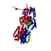
| Movie |
Movie viewer |
|---|---|
| Structure viewer | Molecule: MolmilJmol/JSmol |
- Downloads & links
-Download
| PDBx/mmCIF format | 7kdp.cif.gz | 366.9 KB | Display | PDBx/mmCIF format |
|---|---|---|---|---|
| PDB format | pdb7kdp.ent.gz | 292.3 KB | Display | PDB format |
| PDBx/mmJSON format | 7kdp.json.gz | Tree view | PDBx/mmJSON format | |
| Others |  Other downloads Other downloads |
-Validation report
| Arichive directory | https://data.pdbj.org/pub/pdb/validation_reports/kd/7kdpftp://data.pdbj.org/pub/pdb/validation_reports/kd/7kdp | HTTPS FTP |
|---|
-Related structure data
| Related structure data |  22828MC  7kddC M: map data used to model this data C: citing same article ( |
|---|---|
| Similar structure data |


-Links
 PDBj
PDBj

- Assembly
| Deposited unit | 
|
|---|---|
| 1 |
|
-Components
| #1: Protein | Mass: 102067.688 Da / Num. of mol.: 3 / Source method: isolated from a natural source / Source: (natural) Human cytomegalovirus (strain Towne) / Strain: Towne / References: UniProt: P13201#2: Sugar | ChemComp-NAG /   Type: D-saccharide, beta linking / Mass: 221.208 Da / Num. of mol.: 30 Type: D-saccharide, beta linking / Mass: 221.208 Da / Num. of mol.: 30Source method: isolated from a genetically manipulated source Formula: C8H15NO6 #3: Chemical | 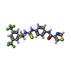  Mass: 518.498 Da / Num. of mol.: 3 Mass: 518.498 Da / Num. of mol.: 3Source method: isolated from a genetically manipulated source Formula: C21H16F6N4OS2 / Feature type: SUBJECT OF INVESTIGATION #4: Chemical | ChemComp-CA / |   Mass: 40.078 Da / Num. of mol.: 1 / Source method: obtained synthetically / Formula: Ca Mass: 40.078 Da / Num. of mol.: 1 / Source method: obtained synthetically / Formula: CaHas ligand of interest | Y | Has protein modification | Y | |
|---|
-Experimental details
-Experiment
| Experiment | Method: ELECTRON MICROSCOPY |
|---|---|
| EM experiment | Aggregation state: PARTICLE / 3D reconstruction method: single particle reconstruction |
- Sample preparation
| Component | Name: HCMV prefusion gB stabilized by a fusion inhibitor / Type: COMPLEX Details: SM5-1 FAb is recombinant expressed with 6xHis and Strep sequence at C-terminal as purification tags. The FAb was used to purify the gB protein from lab cultured HCMV virus (Towne strain)In ...Details: SM5-1 FAb is recombinant expressed with 6xHis and Strep sequence at C-terminal as purification tags. The FAb was used to purify the gB protein from lab cultured HCMV virus (Towne strain)In the density map, the FAb is poorly resolved and not modeled Entity ID: #1 / Source: NATURAL |
|---|---|
| Molecular weight | Value: 0.3 MDa / Experimental value: NO |
| Source (natural) | Organism: Human cytomegalovirus (strain Towne) / Strain: Towne |
| Buffer solution | pH: 7.4 / Details: PBS, 0.1 % DDM, 1 mM EDTA, 2 mg/L WAY-174865 |
| Specimen | Conc.: 0.8 mg/ml / Embedding applied: NO / Shadowing applied: NO / Staining applied: NO / Vitrification applied: YES / Details: this sample was monodispersed |
| Specimen support | Grid material: COPPER / Grid mesh size: 300 divisions/in. / Grid type: Quantifoil R1.2/1.3 |
| Vitrification | Instrument: FEI VITROBOT MARK II / Cryogen name: ETHANE / Humidity: 100 % / Chamber temperature: 277 K / Details: 4 second, -2 force |
- Electron microscopy imaging
| Experimental equipment |  Model: Titan Krios / Image courtesy: FEI Company |
|---|---|
| Microscopy | Model: FEI TITAN KRIOS |
| Electron gun | Electron source:  FIELD EMISSION GUN / Accelerating voltage: 300 kV / Illumination mode: FLOOD BEAM FIELD EMISSION GUN / Accelerating voltage: 300 kV / Illumination mode: FLOOD BEAM |
| Electron lens | Mode: BRIGHT FIELD / Nominal magnification: 18000 X / Nominal defocus max: 2800 nm / Nominal defocus min: 800 nm / Cs: 2.7 mm / C2 aperture diameter: 70 µm / Alignment procedure: COMA FREE |
| Specimen holder | Cryogen: NITROGEN / Specimen holder model: FEI TITAN KRIOS AUTOGRID HOLDER |
| Image recording | Electron dose: 40 e/Å2 / Detector mode: SUPER-RESOLUTION / Film or detector model: GATAN K2 SUMMIT (4k x 4k) / Num. of grids imaged: 3 / Num. of real images: 7768 |
| Image scans | Movie frames/image: 28 / Used frames/image: 1-28 |
- Processing
| Software | Name: PHENIX / Version: dev_3965: / Classification: refinement | ||||||||||||||||||||||||||||||||||||
|---|---|---|---|---|---|---|---|---|---|---|---|---|---|---|---|---|---|---|---|---|---|---|---|---|---|---|---|---|---|---|---|---|---|---|---|---|---|
| EM software |
| ||||||||||||||||||||||||||||||||||||
| Image processing | Details: the movies are motion corrected dose weighted and averaged with and binned 2x in Fourier space with MotionCor2 program | ||||||||||||||||||||||||||||||||||||
| CTF correction | Type: PHASE FLIPPING AND AMPLITUDE CORRECTION | ||||||||||||||||||||||||||||||||||||
| Particle selection | Num. of particles selected: 1906220 Details: a 20 angstrom low pass filtered postfusion structure was used to generate reference images for the particle auto picking | ||||||||||||||||||||||||||||||||||||
| Symmetry | Point symmetry: C3 (3 fold cyclic) | ||||||||||||||||||||||||||||||||||||
| 3D reconstruction | Resolution: 3.6 Å / Resolution method: FSC 0.143 CUT-OFF / Num. of particles: 129837 / Algorithm: BACK PROJECTION / Num. of class averages: 1 / Symmetry type: POINT | ||||||||||||||||||||||||||||||||||||
| Atomic model building | Protocol: FLEXIBLE FIT / Space: REAL | ||||||||||||||||||||||||||||||||||||
| Atomic model building | PDB-ID: 5CXF Accession code: 5CXF / Source name: PDB / Type: experimental model | ||||||||||||||||||||||||||||||||||||
| Refine LS restraints |
|

