Movie
Movie Controller

[English] 日本語
 Yorodumi
- PDB-7kcl: Full-length human mitochondrial Hsp90 (TRAP1) in complex with Sdh... -
+ Open data

- Basic information
| Entry | Database: PDB / ID: 7kcl | |||||||||||||||||||||||||||
|---|---|---|---|---|---|---|---|---|---|---|---|---|---|---|---|---|---|---|---|---|---|---|---|---|---|---|---|---|
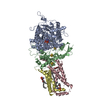
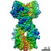
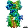
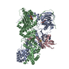
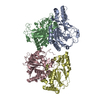
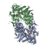
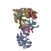
| Title | Full-length human mitochondrial Hsp90 (TRAP1) in complex with SdhB in the presence of AMP-PNP | |||||||||||||||||||||||||||
 Components Components |
| |||||||||||||||||||||||||||
 Keywords Keywords | CHAPERONE / Hsp90 / TRAP1 / SdhB / mitochondria | |||||||||||||||||||||||||||
| Function / homology |  Function and homology information Function and homology informationtranslational attenuation / negative regulation of cellular respiration / Oxidoreductases; Acting on the CH-OH group of donors; With a quinone or similar compound as acceptor / succinate metabolic process / respiratory chain complex II (succinate dehydrogenase) / mitochondrial electron transport, succinate to ubiquinone / Citric acid cycle (TCA cycle) / succinate dehydrogenase (quinone) activity / succinate dehydrogenase / Maturation of TCA enzymes and regulation of TCA cycle ...translational attenuation / negative regulation of cellular respiration / Oxidoreductases; Acting on the CH-OH group of donors; With a quinone or similar compound as acceptor / succinate metabolic process / respiratory chain complex II (succinate dehydrogenase) / mitochondrial electron transport, succinate to ubiquinone / Citric acid cycle (TCA cycle) / succinate dehydrogenase (quinone) activity / succinate dehydrogenase / Maturation of TCA enzymes and regulation of TCA cycle / Respiratory electron transport / tumor necrosis factor receptor binding / 3 iron, 4 sulfur cluster binding / negative regulation of intrinsic apoptotic signaling pathway in response to hydrogen peroxide / ubiquinone binding / proton motive force-driven mitochondrial ATP synthesis / negative regulation of reactive oxygen species biosynthetic process / tricarboxylic acid cycle / aerobic respiration / respiratory electron transport chain / ATP-dependent protein folding chaperone / mitochondrial intermembrane space / 2 iron, 2 sulfur cluster binding / mitochondrial membrane / : / 4 iron, 4 sulfur cluster binding / protein folding / electron transfer activity / mitochondrial inner membrane / mitochondrial matrix / protein kinase binding / ATP hydrolysis activity / mitochondrion / RNA binding / nucleoplasm / ATP binding / membrane / metal ion binding / plasma membrane Similarity search - Function | |||||||||||||||||||||||||||
| Biological species |  Homo sapiens (human) Homo sapiens (human) | |||||||||||||||||||||||||||
| Method | ELECTRON MICROSCOPY / single particle reconstruction / cryo EM / Resolution: 3.14 Å | |||||||||||||||||||||||||||
 Authors Authors | Liu, Y.X. / Agard, D.A. | |||||||||||||||||||||||||||
| Funding support |  United States, 6items United States, 6items
| |||||||||||||||||||||||||||
 Citation Citation | Journal: To Be Published Title: Cryo-EM reveals the dynamic interplay between mitochondrial Hsp90 and SdhB folding intermediates Authors: Liu, Y.X. / Agard, D.A. / Elnatan, D. / Sun, M. / Myasnikov, A.G. | |||||||||||||||||||||||||||
| History |
|
- Structure visualization
| Movie |
Movie viewer |
|---|---|
| Structure viewer | Molecule: MolmilJmol/JSmol |
- Downloads & links
-Download
| PDBx/mmCIF format | 7kcl.cif.gz | 239.6 KB | Display | PDBx/mmCIF format |
|---|---|---|---|---|
| PDB format | pdb7kcl.ent.gz | 188 KB | Display | PDB format |
| PDBx/mmJSON format | 7kcl.json.gz | Tree view | PDBx/mmJSON format | |
| Others |  Other downloads Other downloads |
-Validation report
| Arichive directory | https://data.pdbj.org/pub/pdb/validation_reports/kc/7kclftp://data.pdbj.org/pub/pdb/validation_reports/kc/7kcl | HTTPS FTP |
|---|
-Related structure data
| Related structure data |  22812MC  7kckC  7kcmC M: map data used to model this data C: citing same article ( |
|---|---|
| Similar structure data |


-Links
 PDBj
PDBj


- Assembly
| Deposited unit | 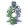
|
|---|---|
| 1 |
|
-Components
| #1: Protein | Mass: 74177.570 Da / Num. of mol.: 2 Source method: isolated from a genetically manipulated source Source: (gene. exp.) Homo sapiens (human) / Gene: TRAP1, HSP75 / Production host:  #2: Protein | | Mass: 15251.708 Da / Num. of mol.: 1 / Fragment: UNP residues 29-160 Source method: isolated from a genetically manipulated source Source: (gene. exp.) Homo sapiens (human) / Gene: SDHB, SDH, SDH1 / Production host: #3: Chemical |   Mass: 506.196 Da / Num. of mol.: 2 / Source method: obtained synthetically / Formula: C10H17N6O12P3 / Comment: AMP-PNP, energy-carrying molecule analogue*YM Mass: 506.196 Da / Num. of mol.: 2 / Source method: obtained synthetically / Formula: C10H17N6O12P3 / Comment: AMP-PNP, energy-carrying molecule analogue*YM#4: Chemical |   Mass: 24.305 Da / Num. of mol.: 2 / Source method: obtained synthetically / Formula: Mg Mass: 24.305 Da / Num. of mol.: 2 / Source method: obtained synthetically / Formula: Mg#5: Chemical |   Mass: 39.098 Da / Num. of mol.: 2 / Source method: obtained synthetically / Formula: K Mass: 39.098 Da / Num. of mol.: 2 / Source method: obtained synthetically / Formula: KHas ligand of interest | N | Has protein modification | N | |
|---|
-Experimental details
-Experiment
| Experiment | Method: ELECTRON MICROSCOPY |
|---|---|
| EM experiment | Aggregation state: PARTICLE / 3D reconstruction method: single particle reconstruction |
- Sample preparation
| Component | Name: human Trap1 in complex with SdhB in the presence of AMP-PNP Type: COMPLEX / Entity ID: #1-#2 / Source: RECOMBINANT |
|---|---|
| Molecular weight | Value: 0.162 MDa / Experimental value: YES |
| Source (natural) | Organism: Homo sapiens (human) |
| Source (recombinant) | Organism: |
| Buffer solution | pH: 7.5 |
| Specimen | Embedding applied: NO / Shadowing applied: NO / Staining applied: NO / Vitrification applied: YES |
| Specimen support | Grid material: COPPER / Grid mesh size: 300 divisions/in. / Grid type: Quantifoil |
| Vitrification | Cryogen name: ETHANE |
- Electron microscopy imaging
| Experimental equipment |  Model: Titan Krios / Image courtesy: FEI Company |
|---|---|
| Microscopy | Model: FEI TITAN KRIOS |
| Electron gun | Electron source:  FIELD EMISSION GUN / Accelerating voltage: 300 kV / Illumination mode: FLOOD BEAM FIELD EMISSION GUN / Accelerating voltage: 300 kV / Illumination mode: FLOOD BEAM |
| Electron lens | Mode: BRIGHT FIELD |
| Image recording | Electron dose: 72 e/Å2 / Detector mode: SUPER-RESOLUTION / Film or detector model: GATAN K2 SUMMIT (4k x 4k) |
- Processing
| Software | Name: PHENIX / Version: 1.18.2_3874: / Classification: refinement | ||||||||||||||||||||||||
|---|---|---|---|---|---|---|---|---|---|---|---|---|---|---|---|---|---|---|---|---|---|---|---|---|---|
| EM software |
| ||||||||||||||||||||||||
| CTF correction | Type: PHASE FLIPPING ONLY | ||||||||||||||||||||||||
| Symmetry | Point symmetry: C1 (asymmetric) | ||||||||||||||||||||||||
| 3D reconstruction | Resolution: 3.14 Å / Resolution method: FSC 0.143 CUT-OFF / Num. of particles: 236804 / Symmetry type: POINT | ||||||||||||||||||||||||
| Atomic model building | Protocol: FLEXIBLE FIT / Space: REAL | ||||||||||||||||||||||||
| Atomic model building | 3D fitting-ID: 1 / Source name: PDB / Type: experimental model
| ||||||||||||||||||||||||
| Refine LS restraints |
|