[English] 日本語
Yorodumi
- PDB-1zoy: Crystal Structure of Mitochondrial Respiratory Complex II from po... -

+
Open data


ID or keywords:

Loading...

-
Basic information

Entry
Database: PDB / ID: 1zoy
Title
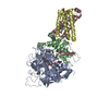
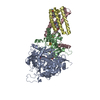
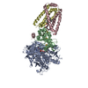
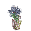
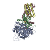
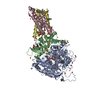
Crystal Structure of Mitochondrial Respiratory Complex II from porcine heart at 2.4 Angstroms
Components
  • FAD-binding protein
  • Iron-sulfur protein
  • Large cytochrome binding protein
  • Small cytochrome binding protein
KeywordsOXIDOREDUCTASE / succinate / ubiquinone oxidoreductase / mitochondrial respiratory Complex II / membrane protein structure
Function / homology
Function and homology information


Citric acid cycle (TCA cycle) / succinate metabolic process / mitochondrial electron transport, succinate to ubiquinone / mitochondrial respiratory chain complex II, succinate dehydrogenase complex (ubiquinone) / succinate dehydrogenase activity / succinate dehydrogenase (quinone) activity / : / succinate dehydrogenase / ubiquinone binding / 3 iron, 4 sulfur cluster binding ...Citric acid cycle (TCA cycle) / succinate metabolic process / mitochondrial electron transport, succinate to ubiquinone / mitochondrial respiratory chain complex II, succinate dehydrogenase complex (ubiquinone) / succinate dehydrogenase activity / succinate dehydrogenase (quinone) activity / : / succinate dehydrogenase / ubiquinone binding / 3 iron, 4 sulfur cluster binding / aerobic respiration / tricarboxylic acid cycle / respiratory electron transport chain / mitochondrial membrane / 2 iron, 2 sulfur cluster binding / flavin adenine dinucleotide binding / nervous system development / 4 iron, 4 sulfur cluster binding / mitochondrial inner membrane / electron transfer activity / heme binding / nucleolus / metal ion binding
Similarity search - Function
Single helix bin / CybS, succinate dehydrogenase cytochrome B small subunit / Succinate dehydrogenase [ubiquinone] cytochrome b small subunit, CybS / succinate dehydrogenase protein domain / Succinate dehydrogenase, cytochrome b556 subunit / Succinate dehydrogenase, cytochrome b subunit, conserved site / Succinate dehydrogenase cytochrome b subunit signature 1. / Succinate dehydrogenase cytochrome b subunit signature 2. / Succinate dehydrogenase, flavoprotein subunit / Fumarate reductase/succinate dehydrogenase, transmembrane subunit ...Single helix bin / CybS, succinate dehydrogenase cytochrome B small subunit / Succinate dehydrogenase [ubiquinone] cytochrome b small subunit, CybS / succinate dehydrogenase protein domain / Succinate dehydrogenase, cytochrome b556 subunit / Succinate dehydrogenase, cytochrome b subunit, conserved site / Succinate dehydrogenase cytochrome b subunit signature 1. / Succinate dehydrogenase cytochrome b subunit signature 2. / Succinate dehydrogenase, flavoprotein subunit / Fumarate reductase/succinate dehydrogenase, transmembrane subunit / Succinate dehydrogenase/fumarate reductase type B, transmembrane subunit / Succinate dehydrogenase/Fumarate reductase transmembrane subunit / Fumarate reductase/succinate dehydrogenase flavoprotein-like, C-terminal domain / Alpha-helical ferredoxin / Fumarate Reductase Iron-sulfur Protein; Chain B, domain 2 / Fumarate reductase/succinate dehydrogenase, transmembrane subunit / Succinate dehydrogenase/fumarate reductase, flavoprotein subunit / Fumarate reductase/succinate dehydrogenase, FAD-binding site / FAD-dependent oxidoreductase SdhA/FrdA/AprA / Fumarate reductase / succinate dehydrogenase FAD-binding site. / 3 helical TM bundles of succinate and fumarate reductases / 4Fe-4S dicluster domain / Flavocytochrome C3; Chain A, domain 1 / Succinate dehydrogenase/fumarate reductase flavoprotein, catalytic domain / Succinate dehydrogenase/fumarate reductase iron-sulphur protein / Succinate dehydogenase/fumarate reductase N-terminal / 2Fe-2S iron-sulfur cluster binding domain / Fumarate reductase/succinate dehydrogenase flavoprotein-like, C-terminal / Fumarate reductase flavoprotein C-term / Fumarate reductase/succinate dehydrogenase flavoprotein-like, C-terminal domain superfamily / Succinate dehydrogenase/fumarate reductase flavoprotein, catalytic domain superfamily / FAD-dependent oxidoreductase 2, FAD binding domain / Alpha-helical ferredoxin / FAD binding domain / Rhinovirus 14, subunit 4 / 2Fe-2S ferredoxin, iron-sulphur binding site / 2Fe-2S ferredoxin-type iron-sulfur binding region signature. / Beta-grasp domain / Beta-grasp domain superfamily / 2Fe-2S ferredoxin-type iron-sulfur binding domain profile. / 2Fe-2S ferredoxin-type iron-sulfur binding domain / 2Fe-2S ferredoxin-like superfamily / 4Fe-4S ferredoxin, iron-sulphur binding, conserved site / 4Fe-4S ferredoxin-type iron-sulfur binding region signature. / 4Fe-4S ferredoxin-type iron-sulfur binding domain profile. / 4Fe-4S ferredoxin-type, iron-sulphur binding domain / FAD/NAD(P)-binding domain / FAD/NAD(P)-binding domain / Methane Monooxygenase Hydroxylase; Chain G, domain 1 / Single alpha-helices involved in coiled-coils or other helix-helix interfaces / 3-Layer(bba) Sandwich / FAD/NAD(P)-binding domain superfamily / Ubiquitin-like (UB roll) / Few Secondary Structures / Irregular / Roll / Alpha-Beta Complex / Up-down Bundle / Orthogonal Bundle / Mainly Alpha / Alpha Beta
Similarity search - Domain/homology
Chem-EPH / FE3-S4 CLUSTER / FLAVIN-ADENINE DINUCLEOTIDE / FE2/S2 (INORGANIC) CLUSTER / PROTOPORPHYRIN IX CONTAINING FE / IRON/SULFUR CLUSTER / UBIQUINONE-1 / Succinate dehydrogenase [ubiquinone] cytochrome b small subunit, mitochondrial / Succinate dehydrogenase cytochrome b560 subunit, mitochondrial / Succinate dehydrogenase [ubiquinone] iron-sulfur subunit, mitochondrial / Succinate dehydrogenase [ubiquinone] flavoprotein subunit, mitochondrial
Similarity search - Component
Biological speciesSus scrofa (pig)
MethodX-RAY DIFFRACTION / SYNCHROTRON / SAD combined with molecular replacement / Resolution: 2.4 Å
AuthorsSun, F. / Huo, X. / Zhai, Y. / Wang, A. / Xu, J. / Su, D. / Bartlam, M. / Rao, Z.
CitationJournal: Cell(Cambridge,Mass.) / Year: 2005
Title: Crystal Structure of Mitochondrial Respiratory Membrane Protein Complex II
Authors: Sun, F. / Huo, X. / Zhai, Y. / Wang, A. / Xu, J. / Su, D. / Bartlam, M. / Rao, Z.
History
DepositionMay 15, 2005Deposition site: RCSB / Processing site: PDBJ
Revision 1.0Jul 12, 2005Provider: repository / Type: Initial release
Revision 1.1Apr 30, 2008Group: Version format compliance
Revision 1.2Jul 13, 2011Group: Version format compliance
Revision 2.0Sep 4, 2019Group: Advisory / Atomic model ...Advisory / Atomic model / Data collection / Derived calculations
Category: atom_site / pdbx_struct_conn_angle ...atom_site / pdbx_struct_conn_angle / pdbx_unobs_or_zero_occ_atoms / struct_conn
Item: _atom_site.B_iso_or_equiv / _atom_site.Cartn_x ..._atom_site.B_iso_or_equiv / _atom_site.Cartn_x / _atom_site.Cartn_y / _atom_site.Cartn_z / _pdbx_struct_conn_angle.ptnr1_label_atom_id / _pdbx_struct_conn_angle.ptnr2_label_atom_id / _pdbx_struct_conn_angle.ptnr3_label_atom_id / _pdbx_struct_conn_angle.value / _struct_conn.pdbx_leaving_atom_flag / _struct_conn.ptnr1_label_atom_id
Revision 2.1Oct 25, 2023Group: Advisory / Data collection ...Advisory / Data collection / Database references / Derived calculations / Refinement description
Category: chem_comp_atom / chem_comp_bond ...chem_comp_atom / chem_comp_bond / database_2 / pdbx_initial_refinement_model / pdbx_struct_conn_angle / pdbx_unobs_or_zero_occ_atoms / struct_conn / struct_site
Item: _database_2.pdbx_DOI / _database_2.pdbx_database_accession ..._database_2.pdbx_DOI / _database_2.pdbx_database_accession / _pdbx_struct_conn_angle.ptnr1_auth_comp_id / _pdbx_struct_conn_angle.ptnr1_auth_seq_id / _pdbx_struct_conn_angle.ptnr1_label_asym_id / _pdbx_struct_conn_angle.ptnr1_label_atom_id / _pdbx_struct_conn_angle.ptnr1_label_comp_id / _pdbx_struct_conn_angle.ptnr1_label_seq_id / _pdbx_struct_conn_angle.ptnr2_auth_comp_id / _pdbx_struct_conn_angle.ptnr2_auth_seq_id / _pdbx_struct_conn_angle.ptnr2_label_asym_id / _pdbx_struct_conn_angle.ptnr2_label_atom_id / _pdbx_struct_conn_angle.ptnr2_label_comp_id / _pdbx_struct_conn_angle.ptnr3_auth_comp_id / _pdbx_struct_conn_angle.ptnr3_auth_seq_id / _pdbx_struct_conn_angle.ptnr3_label_asym_id / _pdbx_struct_conn_angle.ptnr3_label_atom_id / _pdbx_struct_conn_angle.ptnr3_label_comp_id / _pdbx_struct_conn_angle.value / _struct_conn.pdbx_dist_value / _struct_conn.ptnr1_auth_comp_id / _struct_conn.ptnr1_auth_seq_id / _struct_conn.ptnr1_label_asym_id / _struct_conn.ptnr1_label_atom_id / _struct_conn.ptnr1_label_comp_id / _struct_conn.ptnr1_label_seq_id / _struct_conn.ptnr2_auth_comp_id / _struct_conn.ptnr2_auth_seq_id / _struct_conn.ptnr2_label_asym_id / _struct_conn.ptnr2_label_atom_id / _struct_conn.ptnr2_label_comp_id / _struct_conn.ptnr2_label_seq_id / _struct_site.pdbx_auth_asym_id / _struct_site.pdbx_auth_comp_id / _struct_site.pdbx_auth_seq_id

-
Structure visualization

Structure viewerMolecule:
MolmilJmol/JSmol

Downloads & links

-
Assembly

Deposited unit
A: FAD-binding protein
B: Iron-sulfur protein
C: Large cytochrome binding protein
D: Small cytochrome binding protein
hetero molecules


Theoretical massNumber of molelcules
Total (without water)127,24212
Polymers123,3464
Non-polymers3,8958
Water5,963331
1


  • Idetical with deposited unit
  • defined by author&software
TypeNameSymmetry operationNumber
identity operation1_555x,y,z1
Buried area19430 Å2
ΔGint-155 kcal/mol
Surface area40150 Å2
MethodPISA
Unit cell
Length a, b, c (Å)70.238, 83.557, 293.896
Angle α, β, γ (deg.)90.00, 90.00, 90.00
Int Tables number19
Space group name H-MP212121

-
Components

-
Protein , 4 types, 4 molecules ABCD

#1: Protein FAD-binding protein / flavoprotein


Mass: 68313.172 Da / Num. of mol.: 1 / Source method: isolated from a natural source / Source: (natural) Sus scrofa (pig) / Tissue: Porcine heart / References: UniProt: Q0QF01, succinate dehydrogenase
#2: Protein Iron-sulfur protein /


Mass: 28792.268 Da / Num. of mol.: 1 / Source method: isolated from a natural source / Source: (natural) Sus scrofa (pig) / Tissue: Porcine heart / References: UniProt: Q007T0, succinate dehydrogenase
#3: Protein Large cytochrome binding protein


Mass: 15304.081 Da / Num. of mol.: 1 / Source method: isolated from a natural source / Source: (natural) Sus scrofa (pig) / Tissue: Porcine heart / References: UniProt: D0VWV4*PLUS
#4: Protein Small cytochrome binding protein


Mass: 10936.758 Da / Num. of mol.: 1 / Source method: isolated from a natural source / Source: (natural) Sus scrofa (pig) / Tissue: Porcine heart / References: UniProt: A5GZW8

-
Non-polymers , 8 types, 339 molecules

#5: Chemical ChemComp-FAD / FLAVIN-ADENINE DINUCLEOTIDE / Flavin adenine dinucleotide


Mass: 785.550 Da / Num. of mol.: 1 / Source method: obtained synthetically / Formula: C27H33N9O15P2 / Comment: FAD*YM
#6: Chemical ChemComp-FES / FE2/S2 (INORGANIC) CLUSTER / Iron–sulfur cluster


Mass: 175.820 Da / Num. of mol.: 1 / Source method: obtained synthetically / Formula: Fe2S2
#7: Chemical ChemComp-SF4 / IRON/SULFUR CLUSTER / Iron–sulfur cluster


Mass: 351.640 Da / Num. of mol.: 1 / Source method: obtained synthetically / Formula: Fe4S4
#8: Chemical ChemComp-F3S / FE3-S4 CLUSTER / Iron–sulfur cluster


Mass: 295.795 Da / Num. of mol.: 1 / Source method: obtained synthetically / Formula: Fe3S4
#9: Chemical ChemComp-UQ1 / UBIQUINONE-1


Mass: 250.290 Da / Num. of mol.: 1 / Source method: obtained synthetically / Formula: C14H18O4
#10: Chemical ChemComp-HEM / PROTOPORPHYRIN IX CONTAINING FE / HEME / Heme B


Mass: 616.487 Da / Num. of mol.: 1 / Source method: obtained synthetically / Formula: C34H32FeN4O4
#11: Chemical ChemComp-EPH / L-ALPHA-PHOSPHATIDYL-BETA-OLEOYL-GAMMA-PALMITOYL-PHOSPHATIDYLETHANOLAMINE / Phosphatidylethanolamine


Mass: 709.933 Da / Num. of mol.: 2 / Source method: obtained synthetically / Formula: C39H68NO8P / Comment: phospholipid*YM
#12: Water ChemComp-HOH / water / Water


Mass: 18.015 Da / Num. of mol.: 331 / Source method: isolated from a natural source / Formula: H2O

-
Experimental details

-
Experiment

ExperimentMethod: X-RAY DIFFRACTION / Number of used crystals: 2

-
Sample preparation

CrystalDensity Matthews: 3.41 Å3/Da / Density % sol: 63.7 %
Crystal growTemperature: 290 K / Method: vapor diffusion, hanging drop / pH: 7.2
Details: n-nonyl-D-maltoside, n-decyl-D-maltoside, HEPES, PEG 4000, 1,6-hexanediol, NaCl, CaCl2, sucrose, pH 7.2, VAPOR DIFFUSION, HANGING DROP, temperature 290.0K

-
Data collection

Diffraction
IDMean temperature (K)Crystal-ID
11001
21001
Diffraction source
SourceSiteBeamlineIDWavelength (Å)
SYNCHROTRONAPS 19-ID11.74101
SYNCHROTRONAPS 19-ID21.0322
Detector
TypeIDDetectorDate
ADSC QUANTUM 3151CCDNov 1, 2004
ADSC QUANTUM 3152CCDFeb 10, 2005
Radiation
IDMonochromatorProtocolMonochromatic (M) / Laue (L)Scattering typeWavelength-ID
1SAGITALLY FOCUSED Si(111)SADMx-ray1
2SAGITALLY FOCUSED Si(111)SINGLE WAVELENGTHMx-ray1
Radiation wavelength
IDWavelength (Å)Relative weight
11.741011
21.03221
ReflectionResolution: 2.4→50 Å / Num. all: 68771 / Num. obs: 60175 / % possible obs: 85.1 % / Observed criterion σ(F): 1 / Observed criterion σ(I): 1 / Redundancy: 9.1 % / Biso Wilson estimate: 69 Å2 / Rmerge(I) obs: 0.125
Reflection shellResolution: 2.4→2.44 Å / Redundancy: 3.6 % / Rmerge(I) obs: 0.501 / Mean I/σ(I) obs: 2.9 / % possible all: 40.5

-
Processing

Software
NameClassification
HKL-2000data collection
SCALEPACKdata scaling
CNSrefinement
HKL-2000data reduction
CNSphasing
RefinementMethod to determine structure: SAD combined with molecular replacement
Starting model: PDB ENTRY 1NEK
Resolution: 2.4→50 Å / Cross valid method: THROUGHOUT / σ(F): 0 / Stereochemistry target values: Engh & Huber
RfactorNum. reflection% reflectionSelection details
Rfree0.259 5757 8.4 %RANDOM
Rwork0.213 ---
all0.232 68771 --
obs0.232 56559 --
Refine analyze
FreeObs
Luzzati coordinate error0.41 Å0.34 Å
Luzzati d res low-5 Å
Luzzati sigma a0.48 Å0.47 Å
Refinement stepCycle: LAST / Resolution: 2.4→50 Å
ProteinNucleic acidLigandSolventTotal
Num. atoms8480 0 213 331 9024
Refine LS restraints
Refine-IDTypeDev ideal
X-RAY DIFFRACTIONc_bond_d0.018
X-RAY DIFFRACTIONc_angle_deg1.81
X-RAY DIFFRACTIONc_dihedral_angle_d21.82
X-RAY DIFFRACTIONc_improper_angle_d4.83
LS refinement shellResolution: 2.4→2.49 Å
RfactorNum. reflection% reflection
Rfree0.381 280 -
Rwork0.373 --
obs-2637 43 %

+
About Yorodumi

-
News

-
Feb 9, 2022. New format data for meta-information of EMDB entries

New format data for meta-information of EMDB entries

  • Version 3 of the EMDB header file is now the official format.
  • The previous official version 1.9 will be removed from the archive.

Related info.:EMDB header

External links:wwPDB to switch to version 3 of the EMDB data model

-
Aug 12, 2020. Covid-19 info

Covid-19 info

URL: https://pdbj.org/emnavi/covid19.php

New page: Covid-19 featured information page in EM Navigator.

Related info.:Covid-19 info / Mar 5, 2020. Novel coronavirus structure data

+
Mar 5, 2020. Novel coronavirus structure data

Novel coronavirus structure data

Related info.:Yorodumi Speices / Aug 12, 2020. Covid-19 info

External links:COVID-19 featured content - PDBj / Molecule of the Month (242):Coronavirus Proteases

+
Jan 31, 2019. EMDB accession codes are about to change! (news from PDBe EMDB page)

EMDB accession codes are about to change! (news from PDBe EMDB page)

  • The allocation of 4 digits for EMDB accession codes will soon come to an end. Whilst these codes will remain in use, new EMDB accession codes will include an additional digit and will expand incrementally as the available range of codes is exhausted. The current 4-digit format prefixed with “EMD-” (i.e. EMD-XXXX) will advance to a 5-digit format (i.e. EMD-XXXXX), and so on. It is currently estimated that the 4-digit codes will be depleted around Spring 2019, at which point the 5-digit format will come into force.
  • The EM Navigator/Yorodumi systems omit the EMD- prefix.

Related info.:Q: What is EMD? / ID/Accession-code notation in Yorodumi/EM Navigator

External links:EMDB Accession Codes are Changing Soon! / Contact to PDBj

+
Jul 12, 2017. Major update of PDB

Major update of PDB

  • wwPDB released updated PDB data conforming to the new PDBx/mmCIF dictionary.
  • This is a major update changing the version number from 4 to 5, and with Remediation, in which all the entries are updated.
  • In this update, many items about electron microscopy experimental information are reorganized (e.g. em_software).
  • Now, EM Navigator and Yorodumi are based on the updated data.

External links:wwPDB Remediation / Enriched Model Files Conforming to OneDep Data Standards Now Available in the PDB FTP Archive

-
Yorodumi

Thousand views of thousand structures

  • Yorodumi is a browser for structure data from EMDB, PDB, SASBDB, etc.
  • This page is also the successor to EM Navigator detail page, and also detail information page/front-end page for Omokage search.
  • The word "yorodu" (or yorozu) is an old Japanese word meaning "ten thousand". "mi" (miru) is to see.

Related info.:EMDB / PDB / SASBDB / Comparison of 3 databanks / Yorodumi Search / Aug 31, 2016. New EM Navigator & Yorodumi / Yorodumi Papers / Jmol/JSmol / Function and homology information / Changes in new EM Navigator and Yorodumi

Read more