 Movie
Movie Controller


+ Open data

- Basic information
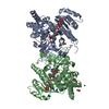
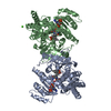
| Entry | Database: PDB / ID: 7k7m | ||||||
|---|---|---|---|---|---|---|---|
| Title | Crystal Structure of a membrane protein | ||||||
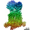
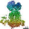
 Components Components | Drug exporters of the RND superfamily-like protein | ||||||
 Keywords Keywords | MEMBRANE PROTEIN / transporter | ||||||
| Function / homology |  Function and homology information Function and homology informationphosphatidylethanolamine transfer activity / phosphatidylglycerol binding / trehalose transmembrane transporter activity / trehalose transport / cell wall biogenesis / mycolate cell wall layer assembly / diacylglycerol binding / cell pole / cell tip / mycolic acid biosynthetic process ...phosphatidylethanolamine transfer activity / phosphatidylglycerol binding / trehalose transmembrane transporter activity / trehalose transport / cell wall biogenesis / mycolate cell wall layer assembly / diacylglycerol binding / cell pole / cell tip / mycolic acid biosynthetic process / phospholipid transport / cell septum / phosphatidylethanolamine binding / cardiolipin binding / phosphatidylinositol binding / regulation of membrane potential / cell wall organization / response to xenobiotic stimulus / response to antibiotic / plasma membrane Similarity search - Function | ||||||
| Biological species |  Mycolicibacterium smegmatis (bacteria) Mycolicibacterium smegmatis (bacteria) | ||||||
| Method |  X-RAY DIFFRACTION / SYNCHROTRON / MOLECULAR REPLACEMENT / Resolution: 3.33 Å X-RAY DIFFRACTION / SYNCHROTRON / MOLECULAR REPLACEMENT / Resolution: 3.33 Å | ||||||
 Authors Authors | Su, C.-C. | ||||||
| Funding support |  United States, 1items United States, 1items
| ||||||
 Citation Citation | Journal: PLoS Biol / Year: 2021 Title: Structures of the mycobacterial membrane protein MmpL3 reveal its mechanism of lipid transport. Authors: Chih-Chia Su / Philip A Klenotic / Meng Cui / Meinan Lyu / Christopher E Morgan / Edward W Yu / Abstract: The mycobacterial membrane protein large 3 (MmpL3) transporter is essential and required for shuttling the lipid trehalose monomycolate (TMM), a precursor of mycolic acid (MA)-containing trehalose ...The mycobacterial membrane protein large 3 (MmpL3) transporter is essential and required for shuttling the lipid trehalose monomycolate (TMM), a precursor of mycolic acid (MA)-containing trehalose dimycolate (TDM) and mycolyl arabinogalactan peptidoglycan (mAGP), in Mycobacterium species, including Mycobacterium tuberculosis and Mycobacterium smegmatis. However, the mechanism that MmpL3 uses to facilitate the transport of fatty acids and lipidic elements to the mycobacterial cell wall remains elusive. Here, we report 7 structures of the M. smegmatis MmpL3 transporter in its unbound state and in complex with trehalose 6-decanoate (T6D) or TMM using single-particle cryo-electron microscopy (cryo-EM) and X-ray crystallography. Combined with calculated results from molecular dynamics (MD) and target MD simulations, we reveal a lipid transport mechanism that involves a coupled movement of the periplasmic domain and transmembrane helices of the MmpL3 transporter that facilitates the shuttling of lipids to the mycobacterial cell wall. | ||||||
| History |
|
- Structure visualization
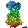
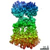
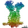
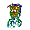
| Structure viewer | Molecule: MolmilJmol/JSmol |
|---|
- Downloads & links
-Download
| PDBx/mmCIF format | 7k7m.cif.gz | 296.2 KB | Display | PDBx/mmCIF format |
|---|---|---|---|---|
| PDB format | pdb7k7m.ent.gz | 239.6 KB | Display | PDB format |
| PDBx/mmJSON format | 7k7m.json.gz | Tree view | PDBx/mmJSON format | |
| Others |  Other downloads Other downloads |
-Validation report
| Arichive directory | https://data.pdbj.org/pub/pdb/validation_reports/k7/7k7mftp://data.pdbj.org/pub/pdb/validation_reports/k7/7k7m | HTTPS FTP |
|---|
-Related structure data
| Related structure data |  7k8aC  7k8bC 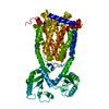 7k8cC  7k8dC  7n6bC 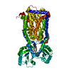 6or2S S: Starting model for refinement C: citing same article ( |
|---|---|
| Similar structure data |


-Links
 PDBj
PDBj- Assembly
| Deposited unit | 
| ||||||||
|---|---|---|---|---|---|---|---|---|---|
| 1 | 
| ||||||||
| 2 | 
| ||||||||
| Unit cell |
|
-Components
| #1: Protein | Mass: 86010.031 Da / Num. of mol.: 2 Source method: isolated from a genetically manipulated source Source: (gene. exp.) Mycolicibacterium smegmatis (strain ATCC 700084 / mc(2)155) (bacteria)Strain: ATCC 700084 / mc(2)155 / Gene: mmpL3, MSMEI_0243 / Production host: #2: Polysaccharide | alpha-D-glucopyranose-(1-1)-6-O-decanoyl-alpha-D-glucopyranose   Type: oligosaccharide, Oligosaccharide / Class: Substrate analog / Mass: 496.546 Da / Num. of mol.: 4 Type: oligosaccharide, Oligosaccharide / Class: Substrate analog / Mass: 496.546 Da / Num. of mol.: 4Source method: isolated from a genetically manipulated source Details: oligosaccharide with reducing-end-to-reducing-end glycosidic bond References: 6-decanoyl-trehalose Has ligand of interest | Y | |
|---|
-Experimental details
-Experiment
| Experiment | Method: X-RAY DIFFRACTION / Number of used crystals: 1 |
|---|
- Sample preparation
| Crystal | Density Matthews: 4.59 Å3/Da / Density % sol: 73.21 % |
|---|---|
| Crystal grow | Temperature: 298 K / Method: vapor diffusion / Details: 20mM Tris, 100mM NaAC and 25% PEG400 |
-Data collection
| Diffraction | Mean temperature: 100 K / Serial crystal experiment: N | |||||||||||||||||||||||||||||||||||||||||||||||||||||||||||||||||||||||||||||||||||||||||||||||||||
|---|---|---|---|---|---|---|---|---|---|---|---|---|---|---|---|---|---|---|---|---|---|---|---|---|---|---|---|---|---|---|---|---|---|---|---|---|---|---|---|---|---|---|---|---|---|---|---|---|---|---|---|---|---|---|---|---|---|---|---|---|---|---|---|---|---|---|---|---|---|---|---|---|---|---|---|---|---|---|---|---|---|---|---|---|---|---|---|---|---|---|---|---|---|---|---|---|---|---|---|---|
| Diffraction source | Source: SYNCHROTRON / Site: APS / Beamline: 24-ID-C / Wavelength: 0.98 Å | |||||||||||||||||||||||||||||||||||||||||||||||||||||||||||||||||||||||||||||||||||||||||||||||||||
| Detector | Type: DECTRIS PILATUS3 S 6M / Detector: PIXEL / Date: Jul 20, 2019 | |||||||||||||||||||||||||||||||||||||||||||||||||||||||||||||||||||||||||||||||||||||||||||||||||||
| Radiation | Protocol: SINGLE WAVELENGTH / Monochromatic (M) / Laue (L): M / Scattering type: x-ray | |||||||||||||||||||||||||||||||||||||||||||||||||||||||||||||||||||||||||||||||||||||||||||||||||||
| Radiation wavelength | Wavelength: 0.98 Å / Relative weight: 1 | |||||||||||||||||||||||||||||||||||||||||||||||||||||||||||||||||||||||||||||||||||||||||||||||||||
| Reflection | Resolution: 3.33→100 Å / Num. obs: 44875 / % possible obs: 99.1 % / Redundancy: 4.6 % / Rmerge(I) obs: 0.089 / Rpim(I) all: 0.046 / Rrim(I) all: 0.101 / Χ2: 0.966 / Net I/σ(I): 5.2 | |||||||||||||||||||||||||||||||||||||||||||||||||||||||||||||||||||||||||||||||||||||||||||||||||||
| Reflection shell | Diffraction-ID: 1
|
- Processing
| Software |
| ||||||||||||||||||||||||||||||||||||||||||||||||||||||||||||||||||||||||||||||||||||||||||||||||
|---|---|---|---|---|---|---|---|---|---|---|---|---|---|---|---|---|---|---|---|---|---|---|---|---|---|---|---|---|---|---|---|---|---|---|---|---|---|---|---|---|---|---|---|---|---|---|---|---|---|---|---|---|---|---|---|---|---|---|---|---|---|---|---|---|---|---|---|---|---|---|---|---|---|---|---|---|---|---|---|---|---|---|---|---|---|---|---|---|---|---|---|---|---|---|---|---|---|
| Refinement | Method to determine structure: MOLECULAR REPLACEMENT Starting model: 6OR2 Resolution: 3.33→98.66 Å / SU ML: 0.72 / Cross valid method: THROUGHOUT / σ(F): 1.34 / Phase error: 41.41 / Stereochemistry target values: ML
| ||||||||||||||||||||||||||||||||||||||||||||||||||||||||||||||||||||||||||||||||||||||||||||||||
| Solvent computation | Shrinkage radii: 0.9 Å / VDW probe radii: 1.11 Å / Solvent model: FLAT BULK SOLVENT MODEL | ||||||||||||||||||||||||||||||||||||||||||||||||||||||||||||||||||||||||||||||||||||||||||||||||
| Displacement parameters | Biso max: 241.42 Å2 / Biso mean: 139.4473 Å2 / Biso min: 82.45 Å2 | ||||||||||||||||||||||||||||||||||||||||||||||||||||||||||||||||||||||||||||||||||||||||||||||||
| Refinement step | Cycle: final / Resolution: 3.33→98.66 Å
| ||||||||||||||||||||||||||||||||||||||||||||||||||||||||||||||||||||||||||||||||||||||||||||||||
| LS refinement shell | Refine-ID: X-RAY DIFFRACTION / Rfactor Rfree error: 0
|
