Method to determine structure: SAD / Resolution: 2.59→99.252 Å / SU ML: 0.51 / Cross valid method: THROUGHOUT / σ(F): 1.33 / Phase error: 36.89 / Stereochemistry target values: ML
Rfactor
Num. reflection
% reflection
Rfree
0.2896
3506
3.87 %
Rwork
0.2492
-
-
obs
0.2507
90546
92.91 %
Solvent computation
Shrinkage radii: 0.9 Å / VDW probe radii: 1.11 Å / Solvent model: FLAT BULK SOLVENT MODEL
Refinement step
Cycle: LAST / Resolution: 2.59→99.252 Å
Protein
Nucleic acid
Ligand
Solvent
Total
Num. atoms
5581
0
85
11
5677
Refine LS restraints
Refine-ID
Type
Dev ideal
Number
X-RAY DIFFRACTION
f_bond_d
0.002
5785
X-RAY DIFFRACTION
f_angle_d
0.523
7857
X-RAY DIFFRACTION
f_dihedral_angle_d
14.564
3464
X-RAY DIFFRACTION
f_chiral_restr
0.038
938
X-RAY DIFFRACTION
f_plane_restr
0.004
982
LS refinement shell
Resolution (Å)
Rfactor Rfree
Num. reflection Rfree
Rfactor Rwork
Num. reflection Rwork
Refine-ID
% reflection obs (%)
2.59-2.6255
0.4669
115
0.4452
2809
X-RAY DIFFRACTION
76
2.6255-2.663
0.4064
111
0.4515
2953
X-RAY DIFFRACTION
79
2.663-2.7028
0.466
131
0.4302
3282
X-RAY DIFFRACTION
88
2.7028-2.745
0.4111
132
0.4019
3359
X-RAY DIFFRACTION
89
2.745-2.79
0.3437
137
0.3821
3548
X-RAY DIFFRACTION
94
2.79-2.8381
0.4059
145
0.3831
3551
X-RAY DIFFRACTION
95
2.8381-2.8897
0.3644
141
0.3656
3602
X-RAY DIFFRACTION
95
2.8897-2.9453
0.3793
148
0.356
3564
X-RAY DIFFRACTION
96
2.9453-3.0054
0.3727
148
0.3554
3590
X-RAY DIFFRACTION
94
3.0054-3.0708
0.3579
142
0.3369
3442
X-RAY DIFFRACTION
93
3.0708-3.1422
0.3382
145
0.3227
3610
X-RAY DIFFRACTION
96
3.1422-3.2208
0.3703
148
0.3139
3643
X-RAY DIFFRACTION
97
3.2208-3.3079
0.3579
150
0.2981
3607
X-RAY DIFFRACTION
97
3.3079-3.4052
0.3029
146
0.274
3606
X-RAY DIFFRACTION
96
3.4052-3.5152
0.3169
149
0.2681
3630
X-RAY DIFFRACTION
96
3.5152-3.6408
0.3502
147
0.2511
3569
X-RAY DIFFRACTION
95
3.6408-3.7866
0.2377
135
0.2289
3435
X-RAY DIFFRACTION
92
3.7866-3.9589
0.2479
147
0.2153
3583
X-RAY DIFFRACTION
96
3.9589-4.1676
0.2464
147
0.2134
3635
X-RAY DIFFRACTION
96
4.1676-4.4288
0.2358
146
0.2001
3576
X-RAY DIFFRACTION
96
4.4288-4.7707
0.2419
140
0.1876
3524
X-RAY DIFFRACTION
94
4.7707-5.2508
0.2283
144
0.1905
3492
X-RAY DIFFRACTION
93
5.2508-6.0105
0.2767
141
0.2379
3551
X-RAY DIFFRACTION
95
6.0105-7.5722
0.2767
135
0.2481
3438
X-RAY DIFFRACTION
92
7.5722-99.3249
0.2945
136
0.2373
3441
X-RAY DIFFRACTION
92
Refinement TLS params.
Method: refined / Refine-ID: X-RAY DIFFRACTION
ID
L11 (°2)
L12 (°2)
L13 (°2)
L22 (°2)
L23 (°2)
L33 (°2)
S11 (Å °)
S12 (Å °)
S13 (Å °)
S21 (Å °)
S22 (Å °)
S23 (Å °)
S31 (Å °)
S32 (Å °)
S33 (Å °)
T11 (Å2)
T12 (Å2)
T13 (Å2)
T22 (Å2)
T23 (Å2)
T33 (Å2)
Origin x (Å)
Origin y (Å)
Origin z (Å)
1
1.2754
-1.0402
-0.6572
2.9095
1.8
4.533
0.0302
-0.0008
0.0573
-0.4234
-0.2859
0.1893
-0.3718
-0.575
0.3061
0.9781
0.0105
0.0217
0.7198
0.0572
0.6637
134.2214
73.662
55.2477
2
2.2657
0.5733
0.1042
1.8868
0.7467
4.3782
0.0084
0.0327
0.0251
0.3934
-0.0216
0.1568
0.81
-0.0046
0.1999
0.8491
-0.0487
0.0523
0.4125
0.0328
0.6567
139.1129
53.4394
32.0505
3
6.854
2.4974
-2.6546
3.1583
-1.2959
2.918
-0.5766
1.8924
-0.1998
-1.6781
0.9354
0.6317
0.3402
-1.4829
-0.1412
1.6891
-0.1845
-0.1775
1.176
-0.2584
0.8933
131.5956
55.2519
12.2247
4
1.1746
-0.3479
-0.6254
1.5199
0.5297
5.1589
0.3472
-0.3787
0.3631
0.0032
-0.0201
-0.5003
-1.4807
0.4836
-0.3053
0.9724
-0.1284
0.0718
0.62
0.0117
0.7601
154.4924
84.8673
51.7131
5
1.3909
1.1242
0.5217
4.2357
3.1991
6.1524
-0.1752
-0.3987
0.0361
-0.2073
0.4625
-0.7
-1.055
1.4035
-0.3056
0.5848
-0.0005
-0.0031
0.9054
0.0405
0.6017
160.4725
74.1595
51.3051
6
2.1226
-0.7247
-1.6245
1.9884
1.5515
7.8949
0.0192
0.2289
0.0129
-0.3943
-0.1152
-0.0086
-0.7906
-0.0451
0.0787
0.6625
-0.0576
-0.1179
0.5186
0.0221
0.533
147.2222
63.2642
21.3636
7
0.0306
-0.0697
0.0926
0.1587
-0.2108
0.2801
0.1581
1.0372
-1.1767
0.1803
-0.1987
-0.6933
0.9437
0.7011
-0.0021
2.6235
0.8053
0.3055
1.5734
-0.4339
1.6751
136.5644
34.8616
8.3098
Refinement TLS group
ID
Refine-ID
Refine TLS-ID
Selection details
1
X-RAY DIFFRACTION
1
chain 'A' and (resid1through207 )
2
X-RAY DIFFRACTION
2
chain 'A' and (resid208through330 )
3
X-RAY DIFFRACTION
3
chain 'A' and (resid331through400 )
4
X-RAY DIFFRACTION
4
chain 'A' and (resid401through501 )
5
X-RAY DIFFRACTION
5
chain 'A' and (resid502through582 )
6
X-RAY DIFFRACTION
6
chain 'A' and (resid583through757 )
7
X-RAY DIFFRACTION
7
chain 'A' and (resid758through761 )
+
About Yorodumi
-
News
-
Feb 9, 2022. New format data for meta-information of EMDB entries
New format data for meta-information of EMDB entries
Version 3 of the EMDB header file is now the official format.
The previous official version 1.9 will be removed from the archive.
In the structure databanks used in Yorodumi, some data are registered as the other names, "COVID-19 virus" and "2019-nCoV". Here are the details of the virus and the list of structure data.
Jan 31, 2019. EMDB accession codes are about to change! (news from PDBe EMDB page)
EMDB accession codes are about to change! (news from PDBe EMDB page)
The allocation of 4 digits for EMDB accession codes will soon come to an end. Whilst these codes will remain in use, new EMDB accession codes will include an additional digit and will expand incrementally as the available range of codes is exhausted. The current 4-digit format prefixed with “EMD-” (i.e. EMD-XXXX) will advance to a 5-digit format (i.e. EMD-XXXXX), and so on. It is currently estimated that the 4-digit codes will be depleted around Spring 2019, at which point the 5-digit format will come into force.
The EM Navigator/Yorodumi systems omit the EMD- prefix.
Related info.:Q: What is EMD? / ID/Accession-code notation in Yorodumi/EM Navigator
Yorodumi is a browser for structure data from EMDB, PDB, SASBDB, etc.
This page is also the successor to EM Navigator detail page, and also detail information page/front-end page for Omokage search.
The word "yorodu" (or yorozu) is an old Japanese word meaning "ten thousand". "mi" (miru) is to see.
Related info.:EMDB / PDB / SASBDB / Comparison of 3 databanks / Yorodumi Search / Aug 31, 2016. New EM Navigator & Yorodumi / Yorodumi Papers / Jmol/JSmol / Function and homology information / Changes in new EM Navigator and Yorodumi
 Movie
Movie Controller

 Yorodumi
 Open data
 Basic information
 Components
 Keywords
 Function and homology information
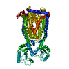
 Mycobacterium smegmatis (bacteria)
 X-RAY DIFFRACTION /
  Authors
 United States, 1items
  Citation
 Structure visualization
 Downloads & links
 Other downloads


 PDBj
PDBj Assembly
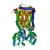
 Mass: 746.050 Da / Num. of mol.: 1 / Source method: obtained synthetically / Formula: C41H80NO8P
 Type: D-saccharide / Mass: 510.615 Da / Num. of mol.: 1 / Source method: obtained synthetically / Formula: C24H46O11 / Comment: detergent*YM
 Mass: 18.015 Da / Num. of mol.: 11 / Source method: isolated from a natural source / Formula: H2O
 Sample preparation
 Processing