 Movie
Movie Controller


+ Open data

- Basic information
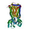
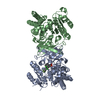
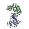
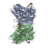
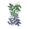
| Entry | Database: PDB / ID: 6n40 | ||||||
|---|---|---|---|---|---|---|---|
| Title | Crystal structure of MmpL3 from Mycobacterium smegmatis | ||||||
 Components Components | Membrane protein, MmpL family protein | ||||||
 Keywords Keywords | MEMBRANE PROTEIN / transporter / ANTIBIOTIC | ||||||
| Function / homology |  Function and homology information Function and homology informationphosphatidylethanolamine transfer activity / phosphatidylglycerol binding / trehalose transmembrane transporter activity / trehalose transport / cell wall biogenesis / mycolate cell wall layer assembly / diacylglycerol binding / cell pole / cell tip / mycolic acid biosynthetic process ...phosphatidylethanolamine transfer activity / phosphatidylglycerol binding / trehalose transmembrane transporter activity / trehalose transport / cell wall biogenesis / mycolate cell wall layer assembly / diacylglycerol binding / cell pole / cell tip / mycolic acid biosynthetic process / phospholipid transport / cell septum / phosphatidylethanolamine binding / cardiolipin binding / phosphatidylinositol binding / regulation of membrane potential / cell wall organization / response to xenobiotic stimulus / response to antibiotic / plasma membrane Similarity search - Function | ||||||
| Biological species |  Mycobacterium smegmatis (bacteria) Mycobacterium smegmatis (bacteria) | ||||||
| Method |  X-RAY DIFFRACTION / SYNCHROTRON / Resolution: 3.307 Å X-RAY DIFFRACTION / SYNCHROTRON / Resolution: 3.307 Å | ||||||
 Authors Authors | Su, C.-C. | ||||||
| Funding support |  United States, 1items United States, 1items
| ||||||
 Citation Citation | Journal: To be published Title: Crystal structure of MmpL3 from Mycobacterium smegmatis Authors: Su, C.-C. / Yu, E.W. | ||||||
| History |
|
- Structure visualization
| Structure viewer | Molecule: MolmilJmol/JSmol |
|---|
- Downloads & links
-Download
| PDBx/mmCIF format | 6n40.cif.gz | 147.8 KB | Display | PDBx/mmCIF format |
|---|---|---|---|---|
| PDB format | pdb6n40.ent.gz | 115.3 KB | Display | PDB format |
| PDBx/mmJSON format | 6n40.json.gz | Tree view | PDBx/mmJSON format | |
| Others |  Other downloads Other downloads |
-Validation report
| Arichive directory | https://data.pdbj.org/pub/pdb/validation_reports/n4/6n40ftp://data.pdbj.org/pub/pdb/validation_reports/n4/6n40 | HTTPS FTP |
|---|
-Related structure data
| Related structure data | |
|---|---|
| Similar structure data |


-Links
 PDBj
PDBj- Assembly
| Deposited unit | 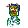
| ||||||||
|---|---|---|---|---|---|---|---|---|---|
| 1 |
| ||||||||
| Unit cell |
|
-Components
| #1: Protein | Mass: 85442.336 Da / Num. of mol.: 1 Source method: isolated from a genetically manipulated source Source: (gene. exp.) Mycobacterium smegmatis (strain ATCC 700084 / mc(2)155) (bacteria)Strain: ATCC 700084 / mc(2)155 / Gene: MSMEG_0250 / Production host: |
|---|
-Experimental details
-Experiment
| Experiment | Method: X-RAY DIFFRACTION / Number of used crystals: 1 |
|---|
- Sample preparation
| Crystal | Density Matthews: 5.02 Å3/Da / Density % sol: 75.51 % |
|---|---|
| Crystal grow | Temperature: 298 K / Method: vapor diffusion, hanging drop / pH: 5 / Details: 20%PEG400 and 0.1M LiCl |
-Data collection
| Diffraction | Mean temperature: 298 K / Serial crystal experiment: N | ||||||||||||||||||||||||||||||||||||||||||||||||||||||||||||||||||||||||||||||||||||||||||||||||||||||||||||||
|---|---|---|---|---|---|---|---|---|---|---|---|---|---|---|---|---|---|---|---|---|---|---|---|---|---|---|---|---|---|---|---|---|---|---|---|---|---|---|---|---|---|---|---|---|---|---|---|---|---|---|---|---|---|---|---|---|---|---|---|---|---|---|---|---|---|---|---|---|---|---|---|---|---|---|---|---|---|---|---|---|---|---|---|---|---|---|---|---|---|---|---|---|---|---|---|---|---|---|---|---|---|---|---|---|---|---|---|---|---|---|---|
| Diffraction source | Source: SYNCHROTRON / Site: APS / Beamline: 24-ID-C / Wavelength: 0.98 Å | ||||||||||||||||||||||||||||||||||||||||||||||||||||||||||||||||||||||||||||||||||||||||||||||||||||||||||||||
| Detector | Type: DECTRIS PILATUS3 S 6M / Detector: PIXEL / Date: Apr 5, 2018 | ||||||||||||||||||||||||||||||||||||||||||||||||||||||||||||||||||||||||||||||||||||||||||||||||||||||||||||||
| Radiation | Protocol: SINGLE WAVELENGTH / Monochromatic (M) / Laue (L): M / Scattering type: x-ray | ||||||||||||||||||||||||||||||||||||||||||||||||||||||||||||||||||||||||||||||||||||||||||||||||||||||||||||||
| Radiation wavelength | Wavelength: 0.98 Å / Relative weight: 1 | ||||||||||||||||||||||||||||||||||||||||||||||||||||||||||||||||||||||||||||||||||||||||||||||||||||||||||||||
| Reflection | Resolution: 3.3→100.185 Å / Num. obs: 26284 / % possible obs: 94 % / Redundancy: 8.4 % / Rmerge(I) obs: 0.145 / Rpim(I) all: 0.054 / Rrim(I) all: 0.155 / Χ2: 1.19 / Net I/σ(I): 5 / Num. measured all: 220075 | ||||||||||||||||||||||||||||||||||||||||||||||||||||||||||||||||||||||||||||||||||||||||||||||||||||||||||||||
| Reflection shell | Diffraction-ID: 1
|
- Processing
| Software |
| ||||||||||||||||||||||||
|---|---|---|---|---|---|---|---|---|---|---|---|---|---|---|---|---|---|---|---|---|---|---|---|---|---|
| Refinement | Resolution: 3.307→100.185 Å / SU ML: 0.56 / Cross valid method: THROUGHOUT / σ(F): 1.33 / Phase error: 43.03 / Stereochemistry target values: ML
| ||||||||||||||||||||||||
| Solvent computation | Shrinkage radii: 0.9 Å / VDW probe radii: 1.11 Å / Solvent model: FLAT BULK SOLVENT MODEL | ||||||||||||||||||||||||
| Refinement step | Cycle: LAST / Resolution: 3.307→100.185 Å
| ||||||||||||||||||||||||
| Refine LS restraints |
|
