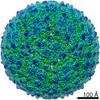
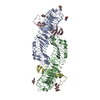
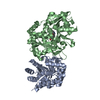
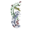
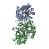
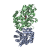
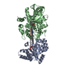
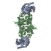
Journal: Nat Commun / Year: 2020 Title: Identification of a pocket factor that is critical to Zika virus assembly. Authors: Nadia M DiNunno / Daniel J Goetschius / Anoop Narayanan / Sydney A Majowicz / Ibrahim Moustafa / Carol M Bator / Susan L Hafenstein / Joyce Jose / Abstract: Zika virus (ZIKV) is an emerging mosquito borne flavivirus and a major public health concern causing severe disease. Due to the presence of a lipid membrane and structural heterogeneity, attaining an ...Zika virus (ZIKV) is an emerging mosquito borne flavivirus and a major public health concern causing severe disease. Due to the presence of a lipid membrane and structural heterogeneity, attaining an atomic resolution structure is challenging, but important to understand virus assembly and life cycle mechanisms that offer distinct targets for therapeutic intervention. We here use subvolume refinement to achieve a 3.4 Å resolution structure and identify two distinct lipid moieties. The first arises from the inner leaflet and is coordinated by hydrophobic residues of the M and E transmembrane helices that form a binding pocket not previously characterized. The second lipid arises from the outer leaflet coordinate between two E protein helices. Structure-based mutagenesis identifies critical hydrophobic interactions and their effect on the virus life cycle. Results show that lipids play an essential role in the ZIKV assembly pathway revealing a potential target of lipid based antiviral drug development.
History
Deposition
Aug 30, 2020
Deposition site: RCSB / Processing site: RCSB
Revision 1.0
Sep 16, 2020
Provider: repository / Type: Initial release
Revision 1.0
Sep 16, 2020
Data content type: EM metadata / Data content type: EM metadata / Provider: repository / Type: Initial release
Revision 1.0
Sep 16, 2020
Data content type: Image / Data content type: Image / Provider: repository / Type: Initial release
Revision 1.0
Sep 16, 2020
Data content type: Primary map / Data content type: Primary map / Provider: repository / Type: Initial release
Revision 1.0
Sep 16, 2020
Data content type: Image / Data content type: Image / Provider: repository / Type: Initial release
Revision 1.0
Sep 16, 2020
Data content type: Primary map / Data content type: Primary map / Provider: repository / Type: Initial release
Revision 1.0
Sep 16, 2020
Data content type: Image / Data content type: Image / Provider: repository / Type: Initial release
Revision 1.0
Sep 16, 2020
Data content type: Primary map / Data content type: Primary map / Provider: repository / Type: Initial release
Data content type: EM metadata / Data content type: EM metadata / EM metadata / Group: Data processing / Experimental summary / Data content type: EM metadata / EM metadata / Category: em_admin / em_software / Data content type: EM metadata / EM metadata / Item: _em_admin.last_update / _em_software.name
In the structure databanks used in Yorodumi, some data are registered as the other names, "COVID-19 virus" and "2019-nCoV". Here are the details of the virus and the list of structure data.
Jan 31, 2019. EMDB accession codes are about to change! (news from PDBe EMDB page)
EMDB accession codes are about to change! (news from PDBe EMDB page)
The allocation of 4 digits for EMDB accession codes will soon come to an end. Whilst these codes will remain in use, new EMDB accession codes will include an additional digit and will expand incrementally as the available range of codes is exhausted. The current 4-digit format prefixed with “EMD-” (i.e. EMD-XXXX) will advance to a 5-digit format (i.e. EMD-XXXXX), and so on. It is currently estimated that the 4-digit codes will be depleted around Spring 2019, at which point the 5-digit format will come into force.
The EM Navigator/Yorodumi systems omit the EMD- prefix.
Related info.:Q: What is EMD? / ID/Accession-code notation in Yorodumi/EM Navigator
Yorodumi is a browser for structure data from EMDB, PDB, SASBDB, etc.
This page is also the successor to EM Navigator detail page, and also detail information page/front-end page for Omokage search.
The word "yorodu" (or yorozu) is an old Japanese word meaning "ten thousand". "mi" (miru) is to see.
Related info.:EMDB / PDB / SASBDB / Comparison of 3 databanks / Yorodumi Search / Aug 31, 2016. New EM Navigator & Yorodumi / Yorodumi Papers / Jmol/JSmol / Function and homology information / Changes in new EM Navigator and Yorodumi
 Movie
Movie Controller


 Open data
 Basic information
 Components
 Keywords
 Function and homology information
 Zika virus
 Authors
 Citation
 Structure visualization
 Downloads & links
 Other downloads


 PDBj
PDBj


 Assembly
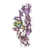
 Sample preparation
 Electron microscopy imaging
 FIELD EMISSION GUN / Accelerating voltage: 300 kV / Illumination mode: FLOOD BEAM
 Processing