 Movie
Movie Controller


+ Open data

- Basic information
| Entry | Database: EMDB / ID: EMD-22527 | |||||||||
|---|---|---|---|---|---|---|---|---|---|---|
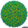
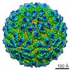
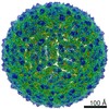
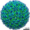
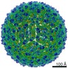
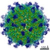
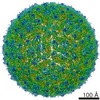
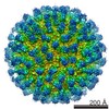
| Title | Icosahedral Map of ZIKV MR-766 | |||||||||
 Map data Map data | Icosahedral Map of ZIKV MR-766 | |||||||||
 Sample Sample |
| |||||||||
| Biological species |   Zika virus Zika virus | |||||||||
| Method | single particle reconstruction / cryo EM / negative staining / Resolution: 3.4 Å | |||||||||
 Authors Authors | Jose J / Hafenstein S | |||||||||
 Citation Citation | Journal: Nat Commun / Year: 2020 Title: Identification of a pocket factor that is critical to Zika virus assembly. Authors: Nadia M DiNunno / Daniel J Goetschius / Anoop Narayanan / Sydney A Majowicz / Ibrahim Moustafa / Carol M Bator / Susan L Hafenstein / Joyce Jose /  Abstract: Zika virus (ZIKV) is an emerging mosquito borne flavivirus and a major public health concern causing severe disease. Due to the presence of a lipid membrane and structural heterogeneity, attaining an ...Zika virus (ZIKV) is an emerging mosquito borne flavivirus and a major public health concern causing severe disease. Due to the presence of a lipid membrane and structural heterogeneity, attaining an atomic resolution structure is challenging, but important to understand virus assembly and life cycle mechanisms that offer distinct targets for therapeutic intervention. We here use subvolume refinement to achieve a 3.4 Å resolution structure and identify two distinct lipid moieties. The first arises from the inner leaflet and is coordinated by hydrophobic residues of the M and E transmembrane helices that form a binding pocket not previously characterized. The second lipid arises from the outer leaflet coordinate between two E protein helices. Structure-based mutagenesis identifies critical hydrophobic interactions and their effect on the virus life cycle. Results show that lipids play an essential role in the ZIKV assembly pathway revealing a potential target of lipid based antiviral drug development. | |||||||||
| History |
|
- Structure visualization
| Movie |
 Movie viewer Movie viewer |
|---|---|
| Structure viewer | EM map: SurfViewMolmilJmol/JSmol |
| Supplemental images |

- Downloads & links
-EMDB archive
| Map data | emd_22527.map.gz | 1 GB | EMDB map data format | |
|---|---|---|---|---|
| Header (meta data) | emd-22527-v30.xmlemd-22527.xml | 8 KB 8 KB | Display Display | EMDB header |
| Images | 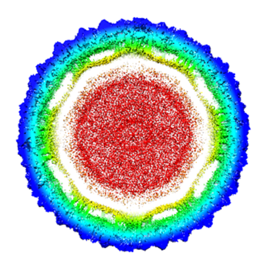 emd_22527.png emd_22527.png | 239.5 KB | ||
| Archive directory |  http://ftp.pdbj.org/pub/emdb/structures/EMD-22527ftp://ftp.pdbj.org/pub/emdb/structures/EMD-22527 http://ftp.pdbj.org/pub/emdb/structures/EMD-22527ftp://ftp.pdbj.org/pub/emdb/structures/EMD-22527 | HTTPS FTP |
-Related structure data


-Links
| EMDB pages | EMDB (EBI/PDBe) / EMDataResource |
|---|---|
| Related items in Molecule of the Month |

-Map
| File | Download / File: emd_22527.map.gz / Format: CCP4 / Size: 1.1 GB / Type: IMAGE STORED AS FLOATING POINT NUMBER (4 BYTES) | ||||||||||||||||||||||||||||||||||||||||||||||||||||||||||||
|---|---|---|---|---|---|---|---|---|---|---|---|---|---|---|---|---|---|---|---|---|---|---|---|---|---|---|---|---|---|---|---|---|---|---|---|---|---|---|---|---|---|---|---|---|---|---|---|---|---|---|---|---|---|---|---|---|---|---|---|---|---|
| Annotation | Icosahedral Map of ZIKV MR-766 | ||||||||||||||||||||||||||||||||||||||||||||||||||||||||||||
| Projections & slices | Image control
Images are generated by Spider. | ||||||||||||||||||||||||||||||||||||||||||||||||||||||||||||
| Voxel size | X=Y=Z: 1.1 Å | ||||||||||||||||||||||||||||||||||||||||||||||||||||||||||||
| Density |
| ||||||||||||||||||||||||||||||||||||||||||||||||||||||||||||
| Symmetry | Space group: 1 | ||||||||||||||||||||||||||||||||||||||||||||||||||||||||||||
| Details | EMDB XML:
CCP4 map header:
| ||||||||||||||||||||||||||||||||||||||||||||||||||||||||||||
 Z (Sec.)
 Y (Row.)
 X (Col.)


-Supplemental data
- Sample components
-Entire : Zika virus
| Entire | Name: Zika virus |
|---|---|
| Components |
|
-Supramolecule #1: Zika virus
| Supramolecule | Name: Zika virus / type: virus / ID: 1 / Parent: 0 / Details: propagated in HeLa cells / NCBI-ID: 64320 / Sci species name: Zika virus / Sci species strain: MR-766 / Virus type: VIRION / Virus isolate: STRAIN / Virus enveloped: No / Virus empty: No |
|---|---|
| Host (natural) | Organism: mosquito (unknown) |
| Virus shell | Shell ID: 1 / Name: Zika / Diameter: 50.0 Å / T number (triangulation number): 3 |
-Experimental details
-Structure determination
| Method | negative staining, cryo EM |
|---|---|
 Processing Processing | single particle reconstruction |
| Aggregation state | particle |
-Sample preparation
| Concentration | 2 mg/mL |
|---|---|
| Buffer | pH: 7 |
| Staining | Type: NEGATIVE / Material: PTA |
| Vitrification | Cryogen name: ETHANE |
- Electron microscopy
| Microscope | FEI TITAN KRIOS |
|---|---|
| Image recording | Film or detector model: FEI FALCON II (4k x 4k) / Average electron dose: 41.7 e/Å2 |
| Electron beam | Acceleration voltage: 300 kV / Electron source:  FIELD EMISSION GUN FIELD EMISSION GUN |
| Electron optics | Illumination mode: FLOOD BEAM / Imaging mode: BRIGHT FIELD |
| Experimental equipment |  Model: Titan Krios / Image courtesy: FEI Company |
-Image processing
| Final reconstruction | Resolution.type: BY AUTHOR / Resolution: 3.4 Å / Resolution method: FSC 0.143 CUT-OFF / Number images used: 9100 |
|---|---|
| Initial angle assignment | Type: MAXIMUM LIKELIHOOD |
| Final angle assignment | Type: MAXIMUM LIKELIHOOD |
