 Movie
Movie Controller

[English] 日本語
 Yorodumi
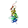
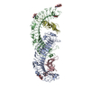
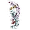
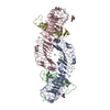
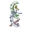
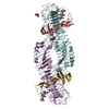
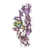
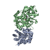
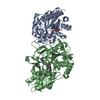
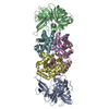
- PDB-4bst: Structure of the ectodomain of LGR5 in complex with R-spondin-1 (... -
+ Open data

- Basic information
| Entry | Database: PDB / ID: 4bst | |||||||||
|---|---|---|---|---|---|---|---|---|---|---|
| Title | Structure of the ectodomain of LGR5 in complex with R-spondin-1 (Fu1Fu2) in P6122 crystal form | |||||||||
 Components Components |
| |||||||||
 Keywords Keywords | SIGNALING PROTEIN / ADULT STEM CELL / LEUCINE-RICH REPEAT G-PROTEIN COUPLED RECEPTOR / LEUCINE-RICH REPEAT / FURIN DOMAIN / WNT SIGNALING / CONGENITAL ANONYCHIA | |||||||||
| Function / homology |  Function and homology information Function and homology informationoocyte differentiation / epithelial cell proliferation involved in renal tubule morphogenesis / protein-hormone receptor activity / regulation of receptor internalization / inner ear development / hair follicle development / positive regulation of Wnt signaling pathway / Regulation of FZD by ubiquitination / trans-Golgi network membrane / positive regulation of protein phosphorylation ...oocyte differentiation / epithelial cell proliferation involved in renal tubule morphogenesis / protein-hormone receptor activity / regulation of receptor internalization / inner ear development / hair follicle development / positive regulation of Wnt signaling pathway / Regulation of FZD by ubiquitination / trans-Golgi network membrane / positive regulation of protein phosphorylation / G protein-coupled receptor binding / G protein-coupled receptor activity / Wnt signaling pathway / transmembrane signaling receptor activity / positive regulation of canonical Wnt signaling pathway / regulation of cell population proliferation / heparin binding / G protein-coupled receptor signaling pathway / signaling receptor binding / : / extracellular region / nucleus / plasma membrane Similarity search - Function | |||||||||
| Biological species |  HOMO SAPIENS (human) HOMO SAPIENS (human) | |||||||||
| Method |  X-RAY DIFFRACTION / SYNCHROTRON / OTHER / Resolution: 4.3 Å X-RAY DIFFRACTION / SYNCHROTRON / OTHER / Resolution: 4.3 Å | |||||||||
 Authors Authors | Peng, W.C. / de Lau, W. / Forneris, F. / Granneman, J.C.M. / Huch, M. / Clevers, H. / Gros, P. | |||||||||
 Citation Citation | Journal: Cell Rep. / Year: 2013 Title: Structure of Stem Cell Growth Factor R-Spondin 1 in Complex with the Ectodomain of its Receptor Lgr5. Authors: Peng, W.C. / De Lau, W. / Forneris, F. / Granneman, J.C.M. / Huch, M. / Clevers, H. / Gros, P. | |||||||||
| History |
|
- Structure visualization
| Structure viewer | Molecule: MolmilJmol/JSmol |
|---|
- Downloads & links
-Download
| PDBx/mmCIF format | 4bst.cif.gz | 466.8 KB | Display | PDBx/mmCIF format |
|---|---|---|---|---|
| PDB format | pdb4bst.ent.gz | 386.2 KB | Display | PDB format |
| PDBx/mmJSON format | 4bst.json.gz | Tree view | PDBx/mmJSON format | |
| Others |  Other downloads Other downloads |
-Validation report
| Arichive directory | https://data.pdbj.org/pub/pdb/validation_reports/bs/4bstftp://data.pdbj.org/pub/pdb/validation_reports/bs/4bst | HTTPS FTP |
|---|
-Related structure data


-Links
 PDBj
PDBj


- Assembly
| Deposited unit | 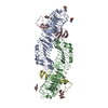
| |||||||||
|---|---|---|---|---|---|---|---|---|---|---|
| 1 |
| |||||||||
| Unit cell |
| |||||||||
| Noncrystallographic symmetry (NCS) | NCS domain:
|
-Components
| #1: Protein | Mass: 60366.457 Da / Num. of mol.: 2 / Fragment: EXTRACELLULAR LRR DOMAIN, RESIDUES 22-543 Source method: isolated from a genetically manipulated source Details: N-LINKED GLYCOSYLATIONS AT ASN 63,77,208,500 / Source: (gene. exp.) HOMO SAPIENS (human) / Cell line (production host): HEK293 / Production host: HOMO SAPIENS (human) / References: UniProt: O75473#2: Protein | Mass: 13706.780 Da / Num. of mol.: 2 / Fragment: FU1FU2, RESIDUES 31-146 Source method: isolated from a genetically manipulated source Source: (gene. exp.) HOMO SAPIENS (human) / Cell line (production host): HEK293 / Production host: HOMO SAPIENS (human) / References: UniProt: Q2MKA7#3: Polysaccharide | beta-D-mannopyranose-(1-4)-2-acetamido-2-deoxy-beta-D-glucopyranose-(1-4)-2-acetamido-2-deoxy-beta- ...beta-D-mannopyranose-(1-4)-2-acetamido-2-deoxy-beta-D-glucopyranose-(1-4)-2-acetamido-2-deoxy-beta-D-glucopyranose | Source method: isolated from a genetically manipulated source #4: Sugar | ChemComp-NAG /   Type: D-saccharide, beta linking / Mass: 221.208 Da / Num. of mol.: 4 Type: D-saccharide, beta linking / Mass: 221.208 Da / Num. of mol.: 4Source method: isolated from a genetically manipulated source Formula: C8H15NO6 Has protein modification | Y | Sequence details | N-TERMINAL 6XHISTAG, PLUS ENLYFQGS AND C-TERMINAL AAA INTRODUCED BY CLONING N-TERMINAL GS AND C- ...N-TERMINAL 6XHISTAG, PLUS ENLYFQGS AND C-TERMINAL AAA INTRODUCED | |
|---|
-Experimental details
-Experiment
| Experiment | Method: X-RAY DIFFRACTION |
|---|
- Sample preparation
| Crystal | Density Matthews: 4.45 Å3/Da / Density % sol: 72.38 % / Description: NONE |
|---|
-Data collection
| Diffraction | Mean temperature: 100 K |
|---|---|
| Diffraction source | Source: SYNCHROTRON / Site: SLS  / Beamline: X06SA / Wavelength: 1 / Beamline: X06SA / Wavelength: 1 |
| Detector | Type: DECTRIS PILATUS 6M / Detector: PIXEL |
| Radiation | Protocol: SINGLE WAVELENGTH / Monochromatic (M) / Laue (L): M / Scattering type: x-ray |
| Radiation wavelength | Wavelength: 1 Å / Relative weight: 1 |
| Reflection | Resolution: 4.31→48.3 Å / Num. obs: 19429 / % possible obs: 99.2 % / Observed criterion σ(I): 2.3 / Redundancy: 7.4 % / Biso Wilson estimate: 177.5 Å2 / Rmerge(I) obs: 0.09 / Net I/σ(I): 12.4 |
- Processing
| Software | Name: PHENIX / Version: (PHENIX.REFINE) / Classification: refinement | ||||||||||||||||||||||||||||||||||||||||||||||||||||||||
|---|---|---|---|---|---|---|---|---|---|---|---|---|---|---|---|---|---|---|---|---|---|---|---|---|---|---|---|---|---|---|---|---|---|---|---|---|---|---|---|---|---|---|---|---|---|---|---|---|---|---|---|---|---|---|---|---|---|
| Refinement | Method to determine structure: OTHER Starting model: NONE Resolution: 4.3→48.171 Å / SU ML: 0.5 / σ(F): 1.34 / Phase error: 26.41 / Stereochemistry target values: ML Details: EXTENDED C-TERMINAL REGION MODELLED WITH ZERO OCCUPANCY
| ||||||||||||||||||||||||||||||||||||||||||||||||||||||||
| Solvent computation | Shrinkage radii: 0.9 Å / VDW probe radii: 1.11 Å / Solvent model: FLAT BULK SOLVENT MODEL | ||||||||||||||||||||||||||||||||||||||||||||||||||||||||
| Displacement parameters | Biso mean: 213.4 Å2 | ||||||||||||||||||||||||||||||||||||||||||||||||||||||||
| Refinement step | Cycle: LAST / Resolution: 4.3→48.171 Å
| ||||||||||||||||||||||||||||||||||||||||||||||||||||||||
| Refine LS restraints |
| ||||||||||||||||||||||||||||||||||||||||||||||||||||||||
| LS refinement shell |
| ||||||||||||||||||||||||||||||||||||||||||||||||||||||||
| Refinement TLS params. | Method: refined / Origin x: 41.6137 Å / Origin y: -12.4197 Å / Origin z: 25.3993 Å
| ||||||||||||||||||||||||||||||||||||||||||||||||||||||||
| Refinement TLS group | Selection details: ALL |
