 Movie
Movie Controller

[English] 日本語
 Yorodumi
- PDB-7jug: C-type carbohydrate-recognition domain 4 of the mannose receptor ... -
+ Open data

- Basic information
| Entry | Database: PDB / ID: 7jug | ||||||
|---|---|---|---|---|---|---|---|
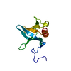
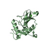
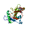
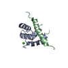
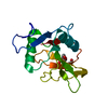
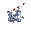
| Title | C-type carbohydrate-recognition domain 4 of the mannose receptor complexed with Man-alpha1-6Man | ||||||
 Components Components | Macrophage mannose receptor 1 | ||||||
 Keywords Keywords | SUGAR BINDING PROTEIN / GLYCOBIOLOGY / CARBOHYDRATE-BINDING PROTEIN / C-TYPE LECTIN / COMPLEX | ||||||
| Function / homology |  Function and homology information Function and homology informationcargo receptor activity / Cross-presentation of soluble exogenous antigens (endosomes) / D-mannose binding / cellular response to interleukin-4 / receptor-mediated endocytosis / cellular response to type II interferon / Modulation by Mtb of host immune system / transmembrane signaling receptor activity / virus receptor activity / cellular response to lipopolysaccharide ...cargo receptor activity / Cross-presentation of soluble exogenous antigens (endosomes) / D-mannose binding / cellular response to interleukin-4 / receptor-mediated endocytosis / cellular response to type II interferon / Modulation by Mtb of host immune system / transmembrane signaling receptor activity / virus receptor activity / cellular response to lipopolysaccharide / signaling receptor activity / endosome membrane / cell surface / plasma membrane Similarity search - Function | ||||||
| Biological species |  Homo sapiens (human) Homo sapiens (human) | ||||||
| Method |  X-RAY DIFFRACTION / SYNCHROTRON / MOLECULAR REPLACEMENT / Resolution: 1.4 Å X-RAY DIFFRACTION / SYNCHROTRON / MOLECULAR REPLACEMENT / Resolution: 1.4 Å | ||||||
 Authors Authors | Weis, W.I. / Feinberg, H. | ||||||
| Funding support |  United Kingdom, 1items United Kingdom, 1items
| ||||||
 Citation Citation | Journal: J.Biol.Chem. / Year: 2021 Title: Structural analysis of carbohydrate binding by the macrophage mannose receptor CD206. Authors: Feinberg, H. / Jegouzo, S.A.F. / Lasanajak, Y. / Smith, D.F. / Drickamer, K. / Weis, W.I. / Taylor, M.E. | ||||||
| History |
|
- Structure visualization
| Structure viewer | Molecule: MolmilJmol/JSmol |
|---|
- Downloads & links
-Download
| PDBx/mmCIF format | 7jug.cif.gz | 77.3 KB | Display | PDBx/mmCIF format |
|---|---|---|---|---|
| PDB format | pdb7jug.ent.gz | 54.7 KB | Display | PDB format |
| PDBx/mmJSON format | 7jug.json.gz | Tree view | PDBx/mmJSON format | |
| Others |  Other downloads Other downloads |
-Validation report
| Arichive directory | https://data.pdbj.org/pub/pdb/validation_reports/ju/7jugftp://data.pdbj.org/pub/pdb/validation_reports/ju/7jug | HTTPS FTP |
|---|
-Related structure data
| Related structure data |  7jubC  7jucC  7judC  7jueC  7jufC  7juhC  7l61C  7l62C  7l63C  7l64C  7l65C 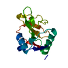 7l66C  7l67C  7l68C C: citing same article ( |
|---|---|
| Similar structure data |


-Links
 PDBj
PDBj


- Assembly
| Deposited unit | 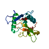
| ||||||||
|---|---|---|---|---|---|---|---|---|---|
| 1 |
| ||||||||
| Unit cell |
|
-Components
| #1: Protein | Mass: 15493.074 Da / Num. of mol.: 1 Source method: isolated from a genetically manipulated source Source: (gene. exp.) Homo sapiens (human) / Gene: MRC1, CLEC13D, CLEC13DL, MRC1L1 / Plasmid: pT5T / Production host:  |
|---|---|
| #2: Chemical | ChemComp-CA /   Mass: 40.078 Da / Num. of mol.: 1 / Source method: obtained synthetically / Formula: Ca Mass: 40.078 Da / Num. of mol.: 1 / Source method: obtained synthetically / Formula: Ca |
| #3: Sugar | ChemComp-MAN /   Type: D-saccharide, alpha linking / Mass: 180.156 Da / Num. of mol.: 1 Type: D-saccharide, alpha linking / Mass: 180.156 Da / Num. of mol.: 1Source method: isolated from a genetically manipulated source Formula: C6H12O6 |
| #4: Water | ChemComp-HOH /  Mass: 18.015 Da / Num. of mol.: 242 / Source method: isolated from a natural source / Formula: H2O Mass: 18.015 Da / Num. of mol.: 242 / Source method: isolated from a natural source / Formula: H2O |
| Has ligand of interest | N |
| Has protein modification | Y |
-Experimental details
-Experiment
| Experiment | Method: X-RAY DIFFRACTION / Number of used crystals: 1 |
|---|
- Sample preparation
| Crystal | Density Matthews: 2.14 Å3/Da / Density % sol: 42.6 % |
|---|---|
| Crystal grow | Temperature: 295 K / Method: vapor diffusion, hanging drop Details: Protein solution: 6.0mg/ml protein in 5mM CaCl2, 10mM Tris, pH 8.0, 25mM NaCl, and 30mM Man-alpha1-6-Man; Reservoir solution: 5% Peg 8K, 0.1M Mes pH=6.0 |
-Data collection
| Diffraction | Mean temperature: 100 K / Serial crystal experiment: N | ||||||||||||||||||||||||||||||
|---|---|---|---|---|---|---|---|---|---|---|---|---|---|---|---|---|---|---|---|---|---|---|---|---|---|---|---|---|---|---|---|
| Diffraction source | Source: SYNCHROTRON / Site: SSRL  / Beamline: BL12-2 / Wavelength: 0.97946 Å / Beamline: BL12-2 / Wavelength: 0.97946 Å | ||||||||||||||||||||||||||||||
| Detector | Type: DECTRIS PILATUS 6M / Detector: PIXEL / Date: Nov 20, 2019 | ||||||||||||||||||||||||||||||
| Radiation | Protocol: SINGLE WAVELENGTH / Monochromatic (M) / Laue (L): M / Scattering type: x-ray | ||||||||||||||||||||||||||||||
| Radiation wavelength | Wavelength: 0.97946 Å / Relative weight: 1 | ||||||||||||||||||||||||||||||
| Reflection | Resolution: 1.4→35.8 Å / Num. obs: 25400 / % possible obs: 98.2 % / Redundancy: 6.8 % / CC1/2: 0.999 / Rmerge(I) obs: 0.04 / Rpim(I) all: 0.016 / Rrim(I) all: 0.044 / Net I/σ(I): 28.1 | ||||||||||||||||||||||||||||||
| Reflection shell | Diffraction-ID: 1
|
- Processing
| Software |
| ||||||||||||||||||||||||||||||||||||||||||||||||||||||||||||
|---|---|---|---|---|---|---|---|---|---|---|---|---|---|---|---|---|---|---|---|---|---|---|---|---|---|---|---|---|---|---|---|---|---|---|---|---|---|---|---|---|---|---|---|---|---|---|---|---|---|---|---|---|---|---|---|---|---|---|---|---|---|
| Refinement | Method to determine structure: MOLECULAR REPLACEMENT Starting model: partially refined model from a different complex Resolution: 1.4→35.8 Å / SU ML: 0.1 / Cross valid method: THROUGHOUT / σ(F): 1.42 / Phase error: 15.29 / Stereochemistry target values: ML
| ||||||||||||||||||||||||||||||||||||||||||||||||||||||||||||
| Solvent computation | Shrinkage radii: 0.9 Å / VDW probe radii: 1.11 Å / Solvent model: FLAT BULK SOLVENT MODEL | ||||||||||||||||||||||||||||||||||||||||||||||||||||||||||||
| Displacement parameters | Biso max: 56.12 Å2 / Biso mean: 15.5987 Å2 / Biso min: 5.18 Å2 | ||||||||||||||||||||||||||||||||||||||||||||||||||||||||||||
| Refinement step | Cycle: final / Resolution: 1.4→35.8 Å
| ||||||||||||||||||||||||||||||||||||||||||||||||||||||||||||
| LS refinement shell | Refine-ID: X-RAY DIFFRACTION / Rfactor Rfree error: 0
|
