 Movie
Movie Controller

[English] 日本語
 Yorodumi
- PDB-1h8u: Crystal Structure of the Eosinophil Major Basic Protein at 1.8A: ... -
+ Open data

- Basic information
| Entry | Database: PDB / ID: 1h8u | ||||||
|---|---|---|---|---|---|---|---|
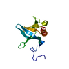
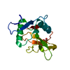
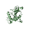
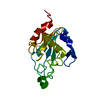
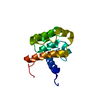
| Title | Crystal Structure of the Eosinophil Major Basic Protein at 1.8A: An Atypical Lectin with a Paradigm Shift in Specificity | ||||||
 Components Components | EOSINOPHIL GRANULE MAJOR BASIC PROTEIN 1 | ||||||
 Keywords Keywords | LECTIN / EOSINOPHIL GRANULE PROTEIN / EMBP | ||||||
| Function / homology |  Function and homology information Function and homology informationextracellular matrix structural constituent conferring compression resistance / defense response to nematode / negative regulation of macrophage cytokine production / negative regulation of interleukin-10 production / positive regulation of interleukin-4 production / transport vesicle / heparin binding / carbohydrate binding / extracellular matrix / ficolin-1-rich granule lumen ...extracellular matrix structural constituent conferring compression resistance / defense response to nematode / negative regulation of macrophage cytokine production / negative regulation of interleukin-10 production / positive regulation of interleukin-4 production / transport vesicle / heparin binding / carbohydrate binding / extracellular matrix / ficolin-1-rich granule lumen / defense response to bacterium / immune response / Neutrophil degranulation / extracellular exosome / extracellular region Similarity search - Function | ||||||
| Biological species |  HOMO SAPIENS (human) HOMO SAPIENS (human) | ||||||
| Method |  X-RAY DIFFRACTION / SYNCHROTRON / MOLECULAR REPLACEMENT / Resolution: 1.8 Å X-RAY DIFFRACTION / SYNCHROTRON / MOLECULAR REPLACEMENT / Resolution: 1.8 Å | ||||||
 Authors Authors | Swaminathan, G.J. / Weaver, A.J. / Loegering, D.A. / Checkel, J.L. / Leonidas, D.D. / Gleich, G.J. / Acharya, K.R. | ||||||
 Citation Citation | Journal: J.Biol.Chem. / Year: 2001 Title: Crystal Structure of the Eosinophil Major Basic Protein at 1.8A. An Atypical Lectin with a Paradigm Shift in Specificity Authors: Swaminathan, G.J. / Weaver, A.J. / Loegering, D.A. / Checkel, J.L. / Leonidas, D.D. / Gleich, G.J. / Acharya, K.R. | ||||||
| History |
|
- Structure visualization
| Structure viewer | Molecule: MolmilJmol/JSmol |
|---|
- Downloads & links
-Download
| PDBx/mmCIF format | 1h8u.cif.gz | 63.5 KB | Display | PDBx/mmCIF format |
|---|---|---|---|---|
| PDB format | pdb1h8u.ent.gz | 47.8 KB | Display | PDB format |
| PDBx/mmJSON format | 1h8u.json.gz | Tree view | PDBx/mmJSON format | |
| Others |  Other downloads Other downloads |
-Validation report
| Arichive directory | https://data.pdbj.org/pub/pdb/validation_reports/h8/1h8uftp://data.pdbj.org/pub/pdb/validation_reports/h8/1h8u | HTTPS FTP |
|---|
-Related structure data
| Related structure data |  1litS S: Starting model for refinement |
|---|---|
| Similar structure data |


-Links
 PDBj
PDBj


- Assembly
| Deposited unit | 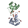
| ||||||||
|---|---|---|---|---|---|---|---|---|---|
| 1 | 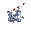
| ||||||||
| 2 | 
| ||||||||
| Unit cell |
| ||||||||
| Noncrystallographic symmetry (NCS) | NCS oper: (Code: given Matrix: (0.717976, -0.022182, 0.695715), Vector: |
-Components
| #1: Protein | Mass: 13820.900 Da / Num. of mol.: 2 / Source method: isolated from a natural source / Source: (natural) HOMO SAPIENS (human) / Cell: EOSINOPHIL / Cellular location: SECRETORY GRANULES / Organelle: PRIMARY GRANULE / Tissue: BLOOD / References: UniProt: P13727#2: Chemical | ChemComp-SO4 /   Mass: 96.063 Da / Num. of mol.: 8 / Source method: obtained synthetically / Formula: SO4 Mass: 96.063 Da / Num. of mol.: 8 / Source method: obtained synthetically / Formula: SO4#3: Chemical |   Mass: 92.094 Da / Num. of mol.: 2 / Source method: obtained synthetically / Formula: C3H8O3 Mass: 92.094 Da / Num. of mol.: 2 / Source method: obtained synthetically / Formula: C3H8O3#4: Water | ChemComp-HOH / |  Mass: 18.015 Da / Num. of mol.: 158 / Source method: isolated from a natural source / Formula: H2O Mass: 18.015 Da / Num. of mol.: 158 / Source method: isolated from a natural source / Formula: H2OCompound details | INVOLVED IN ANTIPARASITIC DEFENSE MECHANISMS AND IMMUNE HYPERSENSITIVITY REACTIONS. INDUCES ...INVOLVED IN ANTIPARASI | Has protein modification | Y | |
|---|
-Experimental details
-Experiment
| Experiment | Method: X-RAY DIFFRACTION / Number of used crystals: 1 |
|---|
- Sample preparation
| Crystal | Density Matthews: 2.32 Å3/Da / Density % sol: 45 % | ||||||||||||||||||||||||||||||
|---|---|---|---|---|---|---|---|---|---|---|---|---|---|---|---|---|---|---|---|---|---|---|---|---|---|---|---|---|---|---|---|
| Crystal grow | pH: 6 Details: 100MM MALONIC ACID PH6, 140MM POTTASIUM DIHYDROGEN PHOSPHATE., pH 6.00 | ||||||||||||||||||||||||||||||
| Crystal grow | *PLUS Temperature: 20 ℃ / Method: vapor diffusion, hanging drop | ||||||||||||||||||||||||||||||
| Components of the solutions | *PLUS
|
-Data collection
| Diffraction | Mean temperature: 100 K |
|---|---|
| Diffraction source | Source: SYNCHROTRON / Site: SRS  / Beamline: PX14.1 / Wavelength: 1.244 / Beamline: PX14.1 / Wavelength: 1.244 |
| Detector | Type: ADSC CCD / Detector: CCD / Date: May 15, 2000 / Details: MIRRORS |
| Radiation | Protocol: SINGLE WAVELENGTH / Monochromatic (M) / Laue (L): M / Scattering type: x-ray |
| Radiation wavelength | Wavelength: 1.244 Å / Relative weight: 1 |
| Reflection | Resolution: 1.8→40 Å / Num. obs: 21127 / % possible obs: 96 % / Redundancy: 10.27 % / Biso Wilson estimate: 24 Å2 / Rsym value: 0.07 / Net I/σ(I): 16.2 |
| Reflection shell | Resolution: 1.8→1.86 Å / Mean I/σ(I) obs: 5.18 / Rsym value: 0.229 / % possible all: 91.8 |
| Reflection | *PLUS Lowest resolution: 40 Å / Num. measured all: 217030 / Rmerge(I) obs: 0.07 |
| Reflection shell | *PLUS % possible obs: 91.8 % / Rmerge(I) obs: 0.229 |
- Processing
| Software |
| ||||||||||||||||||||||||||||||||||||||||||||||||||||||||||||||||||||||||||||||||
|---|---|---|---|---|---|---|---|---|---|---|---|---|---|---|---|---|---|---|---|---|---|---|---|---|---|---|---|---|---|---|---|---|---|---|---|---|---|---|---|---|---|---|---|---|---|---|---|---|---|---|---|---|---|---|---|---|---|---|---|---|---|---|---|---|---|---|---|---|---|---|---|---|---|---|---|---|---|---|---|---|---|
| Refinement | Method to determine structure: MOLECULAR REPLACEMENT Starting model: PDB ENTRY 1LIT Resolution: 1.8→36.18 Å / Rfactor Rfree error: 0.008 / Data cutoff high absF: 905624.99 / Isotropic thermal model: RESTRAINED / Cross valid method: THROUGHOUT / σ(F): 0 Details: N-TERMINAL RESIDUES THR A1 AND CYS A2 WERE NOT SEEN IN ELECTRON DENSITY MAPS N-TERMINAL RESIDUE THR B1 WERE NOT SEEN IN ELECTRON DENSITY MAPS. NO SIDE CHAIN DENSITY SEEN FOR RESIDUES ARG B3 ...Details: N-TERMINAL RESIDUES THR A1 AND CYS A2 WERE NOT SEEN IN ELECTRON DENSITY MAPS N-TERMINAL RESIDUE THR B1 WERE NOT SEEN IN ELECTRON DENSITY MAPS. NO SIDE CHAIN DENSITY SEEN FOR RESIDUES ARG B3 AND ARG B98 WHICH WERE MODELED AS ALANINES IN THE FINAL STRUCTURE.
| ||||||||||||||||||||||||||||||||||||||||||||||||||||||||||||||||||||||||||||||||
| Solvent computation | Solvent model: FLAT MODEL / Bsol: 52.0733 Å2 / ksol: 0.386404 e/Å3 | ||||||||||||||||||||||||||||||||||||||||||||||||||||||||||||||||||||||||||||||||
| Displacement parameters | Biso mean: 27.1 Å2
| ||||||||||||||||||||||||||||||||||||||||||||||||||||||||||||||||||||||||||||||||
| Refine analyze |
| ||||||||||||||||||||||||||||||||||||||||||||||||||||||||||||||||||||||||||||||||
| Refinement step | Cycle: LAST / Resolution: 1.8→36.18 Å
| ||||||||||||||||||||||||||||||||||||||||||||||||||||||||||||||||||||||||||||||||
| Refine LS restraints |
| ||||||||||||||||||||||||||||||||||||||||||||||||||||||||||||||||||||||||||||||||
| LS refinement shell | Resolution: 1.8→1.91 Å / Rfactor Rfree error: 0.031 / Total num. of bins used: 6
| ||||||||||||||||||||||||||||||||||||||||||||||||||||||||||||||||||||||||||||||||
| Xplor file |
| ||||||||||||||||||||||||||||||||||||||||||||||||||||||||||||||||||||||||||||||||
| Software | *PLUS Name: CNS / Version: 1 / Classification: refinement | ||||||||||||||||||||||||||||||||||||||||||||||||||||||||||||||||||||||||||||||||
| Refine LS restraints | *PLUS
| ||||||||||||||||||||||||||||||||||||||||||||||||||||||||||||||||||||||||||||||||
| LS refinement shell | *PLUS Rfactor Rwork: 0.36 |
