[English] 日本語
Yorodumi
- PDB-7jsy: Proteinase K soaked with I3C determined by MicroED from a single ... -

+
Open data


ID or keywords:

Loading...

-
Basic information

Entry
Database: PDB / ID: 7jsy
TitleProteinase K soaked with I3C determined by MicroED from a single milled microcrystal
ComponentsProteinase K
KeywordsHYDROLASE / Proteinase K
Function / homology
Function and homology information


peptidase K / serine-type endopeptidase activity / proteolysis / extracellular region / metal ion binding
Similarity search - Function
Proteinase K-like catalytic domain / Peptidase S8 propeptide/proteinase inhibitor I9 / Peptidase inhibitor I9 / Peptidase S8 propeptide/proteinase inhibitor I9 superfamily / Peptidase S8, subtilisin, His-active site / Serine proteases, subtilase family, histidine active site. / Serine proteases, subtilase family, aspartic acid active site. / Peptidase S8, subtilisin, Asp-active site / Serine proteases, subtilase family, serine active site. / Peptidase S8, subtilisin, Ser-active site ...Proteinase K-like catalytic domain / Peptidase S8 propeptide/proteinase inhibitor I9 / Peptidase inhibitor I9 / Peptidase S8 propeptide/proteinase inhibitor I9 superfamily / Peptidase S8, subtilisin, His-active site / Serine proteases, subtilase family, histidine active site. / Serine proteases, subtilase family, aspartic acid active site. / Peptidase S8, subtilisin, Asp-active site / Serine proteases, subtilase family, serine active site. / Peptidase S8, subtilisin, Ser-active site / Serine proteases, subtilase domain profile. / Peptidase S8, subtilisin-related / Peptidase S8/S53 domain superfamily / Subtilase family / Peptidase S8/S53 domain
Similarity search - Domain/homology
Chem-I3C / Proteinase K
Similarity search - Component
Biological speciesParengyodontium album (fungus)
MethodELECTRON CRYSTALLOGRAPHY / electron crystallography / cryo EM / Resolution: 1.78 Å
AuthorsMartynowycz, M.W. / Gonen, T.
Funding support United States, 2items
OrganizationGrant numberCountry
Howard Hughes Medical Institute (HHMI) United States
National Institutes of Health/National Institute of General Medical Sciences (NIH/NIGMS)P41GM136508 United States
Citation
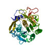
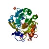
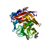
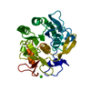
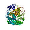
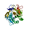
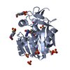
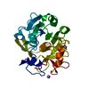
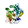
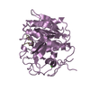
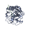
Journal: Structure / Year: 2021
Title: Ligand Incorporation into Protein Microcrystals for MicroED by On-Grid Soaking.
Authors: Michael W Martynowycz / Tamir Gonen /
Abstract: A high throughout method for soaking ligands into protein microcrystals on TEM grids is presented. Every crystal on the grid is soaked simultaneously using only standard cryoelectron microscopy ...A high throughout method for soaking ligands into protein microcrystals on TEM grids is presented. Every crystal on the grid is soaked simultaneously using only standard cryoelectron microscopy vitrification equipment. The method is demonstrated using proteinase K microcrystals soaked with the 5-amino-2,4,6-triodoisophthalic acid (I3C) magic triangle. A soaked microcrystal is milled to a thickness of approximately 200 nm using a focused ion beam, and MicroED data are collected. A high-resolution structure of the protein with four ligands at high occupancy is determined. Both the number of ligands bound and their occupancy is higher using on-grid soaking of microcrystals compared with much larger crystals treated similarly and investigated by X-ray crystallography. These results indicate that on-grid soaking ligands into microcrystals results in efficient uptake of ligands into protein microcrystals.
History
DepositionAug 16, 2020Deposition site: RCSB / Processing site: RCSB
Revision 1.0Oct 14, 2020Provider: repository / Type: Initial release
Revision 1.1Jan 20, 2021Group: Database references / Category: citation
Item: _citation.journal_volume / _citation.page_first / _citation.year

-
Structure visualization

Movie
  • Deposited structure unit
  • Imaged by Jmol
  • Download
  • Simplified surface model + fitted atomic model
  • EMDB-22463
  • Imaged by Jmol
  • Download
  • Superimposition on EM map
  • EMDB-22463
  • Imaged by UCSF Chimera
  • Download
Movie viewer
Structure viewerMolecule:
MolmilJmol/JSmol

Downloads & links

-
Assembly

Deposited unit
A: Proteinase K
hetero molecules


Theoretical massNumber of molelcules
Total (without water)31,1665
Polymers28,9311
Non-polymers2,2354
Water4,432246
1


  • Idetical with deposited unit
  • defined by author
TypeNameSymmetry operationNumber
identity operation1_555x,y,z1
Buried area0 Å2
ΔGint0 kcal/mol
Surface area10650 Å2
Unit cell
Length a, b, c (Å)67.555, 67.555, 102.773
Angle α, β, γ (deg.)90.000, 90.000, 90.000
Int Tables number96
Space group name H-MP43212
Space group name HallP4nw2abw
Symmetry operation#1: x,y,z
#2: -y+1/2,x+1/2,z+3/4
#3: y+1/2,-x+1/2,z+1/4
#4: x+1/2,-y+1/2,-z+1/4
#5: -x+1/2,y+1/2,-z+3/4
#6: -x,-y,z+1/2
#7: y,x,-z
#8: -y,-x,-z+1/2
Components on special symmetry positions
IDModelComponents
11A-703-

HOH

-
Components

#1: Protein Proteinase K / / Endopeptidase K / Tritirachium alkaline proteinase


Mass: 28930.783 Da / Num. of mol.: 1 / Source method: obtained synthetically / Source: (synth.) Parengyodontium album (fungus) / References: UniProt: P06873, peptidase K
#2: Chemical
ChemComp-I3C / 5-amino-2,4,6-triiodobenzene-1,3-dicarboxylic acid / 5-Amino-2,4,6-triiodoisophthalic acid


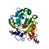
Mass: 558.835 Da / Num. of mol.: 4 / Source method: obtained synthetically / Formula: C8H4I3NO4
#3: Water ChemComp-HOH / water / Water


Mass: 18.015 Da / Num. of mol.: 246 / Source method: isolated from a natural source / Formula: H2O
Has ligand of interestN

-
Experimental details

-
Experiment

ExperimentMethod: ELECTRON CRYSTALLOGRAPHY
EM experimentAggregation state: 3D ARRAY / 3D reconstruction method: electron crystallography

-
Sample preparation

ComponentName: Proteinase K / Type: COMPLEX / Details: Serine protease / Entity ID: #1 / Source: NATURAL
Molecular weightValue: 0.0289 MDa / Experimental value: NO
Source (natural)Organism: Parengyodontium album (fungus)
Buffer solutionpH: 7.5
SpecimenConc.: 5 mg/ml / Embedding applied: NO / Shadowing applied: NO / Staining applied: NO / Vitrification applied: YES
Specimen supportGrid material: COPPER / Grid mesh size: 200 divisions/in. / Grid type: Quantifoil R2/2
VitrificationCryogen name: ETHANE

-
Data collection

MicroscopyModel: TFS TALOS
Details: Tilt series: -30 to +30, 0.25 deg/sec, 1 sec readout
Electron gunElectron source: FIELD EMISSION GUN / Accelerating voltage: 200 kV / Illumination mode: FLOOD BEAM
Electron lensMode: DIFFRACTION / Nominal defocus min: 0 nm / C2 aperture diameter: 70 µm / Alignment procedure: BASIC
Specimen holderCryogen: NITROGEN / Specimen holder model: FEI TITAN KRIOS AUTOGRID HOLDER / Temperature (min): 70 K
Image recordingAverage exposure time: 1 sec. / Electron dose: 0.01 e/Å2 / Film or detector model: FEI CETA (4k x 4k) / Num. of diffraction images: 240 / Num. of grids imaged: 1 / Num. of real images: 240
Image scansSampling size: 28 µm / Width: 4096 / Height: 4096
EM diffractionCamera length: 1900 mm / Tilt angle list: -30,30
EM diffraction shellResolution: 43.32→1.78 Å / Fourier space coverage: 94.52 % / Multiplicity: 4.9 / Num. of structure factors: 10458 / Phase residual: 18 °
EM diffraction statsFourier space coverage: 94.52 % / High resolution: 1.78 Å / Num. of intensities measured: 109326 / Num. of structure factors: 10458 / Phase error: 18 ° / Phase residual: 18 ° / Phase error rejection criteria: 0 / Rmerge: 0.26 / Rsym: 0.13
ReflectionBiso Wilson estimate: 14.37 Å2

-
Processing

Software
NameVersionClassificationNB
phenix.refine1.17.1_3660refinement
PHENIX1.17.1_3660refinement
EM 3D crystal entity∠α: 90 ° / ∠β: 90 ° / ∠γ: 90 ° / A: 67.55 Å / B: 67.55 Å / C: 102.77 Å / Space group name: 96 / Space group num: 96
CTF correctionType: NONE
3D reconstructionResolution: 1.78 Å / Resolution method: DIFFRACTION PATTERN/LAYERLINES / Algorithm: FOURIER SPACE / Symmetry type: 3D CRYSTAL
Atomic model buildingB value: 13.51 / Protocol: RIGID BODY FIT / Space: RECIPROCAL / Target criteria: maximum liklihood
Atomic model buildingPDB-ID: 6CL7
Pdb chain-ID: A / Pdb chain residue range: 1-279
RefinementResolution: 1.78→43.32 Å / SU ML: 0.2175 / Cross valid method: FREE R-VALUE / σ(F): 1.34 / Phase error: 19.6251
Stereochemistry target values: GeoStd + Monomer Library + CDL v1.2
RfactorNum. reflection% reflection
Rfree0.2094 1449 6.53 %
Rwork0.1694 20756 -
obs0.1721 22205 94.52 %
Solvent computationShrinkage radii: 0.9 Å / VDW probe radii: 1.11 Å / Solvent model: FLAT BULK SOLVENT MODEL
Displacement parametersBiso mean: 13.51 Å2
Refinement stepCycle: LAST / Resolution: 1.78→43.32 Å
ProteinNucleic acidLigandSolventTotal
Num. atoms2093 0 0 246 2339
Refine LS restraints
Refine-IDTypeDev idealNumber
ELECTRON CRYSTALLOGRAPHYf_bond_d0.00482132
ELECTRON CRYSTALLOGRAPHYf_angle_d0.76312906
ELECTRON CRYSTALLOGRAPHYf_chiral_restr0.045312
ELECTRON CRYSTALLOGRAPHYf_plane_restr0.0038382
ELECTRON CRYSTALLOGRAPHYf_dihedral_angle_d6.8398315
LS refinement shell
Resolution (Å)Rfactor RfreeNum. reflection RfreeRfactor RworkNum. reflection RworkRefine-ID% reflection obs (%)
1.78-1.840.35171400.28792033ELECTRON CRYSTALLOGRAPHY94.56
1.84-1.920.26051400.22852039ELECTRON CRYSTALLOGRAPHY95.15
1.92-20.25731540.19392037ELECTRON CRYSTALLOGRAPHY94.97
2-2.110.26311470.18312043ELECTRON CRYSTALLOGRAPHY94.93
2.11-2.240.22881360.17092059ELECTRON CRYSTALLOGRAPHY94.73
2.24-2.420.22541480.15512068ELECTRON CRYSTALLOGRAPHY94.62
2.42-2.660.22371460.16512049ELECTRON CRYSTALLOGRAPHY94.29
2.66-3.040.17771430.15642102ELECTRON CRYSTALLOGRAPHY94.61
3.04-3.830.16431410.14592103ELECTRON CRYSTALLOGRAPHY94.13

+
About Yorodumi

-
News

-
Feb 9, 2022. New format data for meta-information of EMDB entries

New format data for meta-information of EMDB entries

  • Version 3 of the EMDB header file is now the official format.
  • The previous official version 1.9 will be removed from the archive.

Related info.:EMDB header

External links:wwPDB to switch to version 3 of the EMDB data model

-
Aug 12, 2020. Covid-19 info

Covid-19 info

URL: https://pdbj.org/emnavi/covid19.php

New page: Covid-19 featured information page in EM Navigator.

Related info.:Covid-19 info / Mar 5, 2020. Novel coronavirus structure data

+
Mar 5, 2020. Novel coronavirus structure data

Novel coronavirus structure data

Related info.:Yorodumi Speices / Aug 12, 2020. Covid-19 info

External links:COVID-19 featured content - PDBj / Molecule of the Month (242):Coronavirus Proteases

+
Jan 31, 2019. EMDB accession codes are about to change! (news from PDBe EMDB page)

EMDB accession codes are about to change! (news from PDBe EMDB page)

  • The allocation of 4 digits for EMDB accession codes will soon come to an end. Whilst these codes will remain in use, new EMDB accession codes will include an additional digit and will expand incrementally as the available range of codes is exhausted. The current 4-digit format prefixed with “EMD-” (i.e. EMD-XXXX) will advance to a 5-digit format (i.e. EMD-XXXXX), and so on. It is currently estimated that the 4-digit codes will be depleted around Spring 2019, at which point the 5-digit format will come into force.
  • The EM Navigator/Yorodumi systems omit the EMD- prefix.

Related info.:Q: What is EMD? / ID/Accession-code notation in Yorodumi/EM Navigator

External links:EMDB Accession Codes are Changing Soon! / Contact to PDBj

+
Jul 12, 2017. Major update of PDB

Major update of PDB

  • wwPDB released updated PDB data conforming to the new PDBx/mmCIF dictionary.
  • This is a major update changing the version number from 4 to 5, and with Remediation, in which all the entries are updated.
  • In this update, many items about electron microscopy experimental information are reorganized (e.g. em_software).
  • Now, EM Navigator and Yorodumi are based on the updated data.

External links:wwPDB Remediation / Enriched Model Files Conforming to OneDep Data Standards Now Available in the PDB FTP Archive

-
Yorodumi

Thousand views of thousand structures

  • Yorodumi is a browser for structure data from EMDB, PDB, SASBDB, etc.
  • This page is also the successor to EM Navigator detail page, and also detail information page/front-end page for Omokage search.
  • The word "yorodu" (or yorozu) is an old Japanese word meaning "ten thousand". "mi" (miru) is to see.

Related info.:EMDB / PDB / SASBDB / Comparison of 3 databanks / Yorodumi Search / Aug 31, 2016. New EM Navigator & Yorodumi / Yorodumi Papers / Jmol/JSmol / Function and homology information / Changes in new EM Navigator and Yorodumi

Read more