 Movie
Movie Controller

[English] 日本語
 Yorodumi
- PDB-7jlz: Crystal structure of 30S ribosomal A1408 methyltransferase from a... -
+ Open data

- Basic information
| Entry | Database: PDB / ID: 7jlz | ||||||
|---|---|---|---|---|---|---|---|
| Title | Crystal structure of 30S ribosomal A1408 methyltransferase from an uncultured bacterium (UncKam) | ||||||
 Components Components | 16S rRNA methylase | ||||||
 Keywords Keywords | RNA BINDING PROTEIN / Ribosome methylation / Aminoglycoside resistance / A1408 methyltransferase | ||||||
| Function / homology | tRNA (guanine(46)-N7)-methyltransferase activity / tRNA (guanine-N-7) methyltransferase, Trmb type / Putative methyltransferase / S-adenosyl-L-methionine-dependent methyltransferase superfamily / 16S rRNA methylase Function and homology information Function and homology information | ||||||
| Biological species |  uncultured bacterium (environmental samples) uncultured bacterium (environmental samples) | ||||||
| Method |  X-RAY DIFFRACTION / SYNCHROTRON / MOLECULAR REPLACEMENT / Resolution: 1.64 Å X-RAY DIFFRACTION / SYNCHROTRON / MOLECULAR REPLACEMENT / Resolution: 1.64 Å | ||||||
 Authors Authors | Nosrati, M. / Conn, G.L. | ||||||
| Funding support |  United States, 1items United States, 1items
| ||||||
 Citation Citation | Journal: To be published Title: Roles of conserved tryptophans in substrate recognition and catalysis by the aminoglycoside-resistance 16S rRNA (m1A1408) rRNA methyltransferases Authors: Nosrati, M. / Witek, M.A. / Dayne, W. / Zelinskaya, N. / Dunham, C.M. / Conn, G.L. | ||||||
| History |
|
- Structure visualization
| Structure viewer | Molecule: MolmilJmol/JSmol |
|---|
- Downloads & links
-Download
| PDBx/mmCIF format | 7jlz.cif.gz | 104.5 KB | Display | PDBx/mmCIF format |
|---|---|---|---|---|
| PDB format | pdb7jlz.ent.gz | 79.2 KB | Display | PDB format |
| PDBx/mmJSON format | 7jlz.json.gz | Tree view | PDBx/mmJSON format | |
| Others |  Other downloads Other downloads |
-Validation report
| Arichive directory | https://data.pdbj.org/pub/pdb/validation_reports/jl/7jlzftp://data.pdbj.org/pub/pdb/validation_reports/jl/7jlz | HTTPS FTP |
|---|
-Related structure data
| Related structure data |  3mteS S: Starting model for refinement |
|---|---|
| Similar structure data |


-Links
 PDBj
PDBj

- Assembly
| Deposited unit | 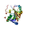
| |||||||||
|---|---|---|---|---|---|---|---|---|---|---|
| 1 |
| |||||||||
| Unit cell |
| |||||||||
| Components on special symmetry positions |
|
-Components
| #1: Protein | Mass: 24403.965 Da / Num. of mol.: 1 Source method: isolated from a genetically manipulated source Source: (gene. exp.) uncultured bacterium (environmental samples)Gene: ACD_24C00409G0003 / Production host: | ||||
|---|---|---|---|---|---|
| #2: Chemical | ChemComp-SO4 /   Mass: 96.063 Da / Num. of mol.: 5 / Source method: obtained synthetically / Formula: SO4 Mass: 96.063 Da / Num. of mol.: 5 / Source method: obtained synthetically / Formula: SO4#3: Water | ChemComp-HOH / |  Mass: 18.015 Da / Num. of mol.: 126 / Source method: isolated from a natural source / Formula: H2O Mass: 18.015 Da / Num. of mol.: 126 / Source method: isolated from a natural source / Formula: H2OHas ligand of interest | N | |
-Experimental details
-Experiment
| Experiment | Method: X-RAY DIFFRACTION / Number of used crystals: 1 |
|---|
- Sample preparation
| Crystal | Density Matthews: 2.28 Å3/Da / Density % sol: 45.96 % |
|---|---|
| Crystal grow | Temperature: 277 K / Method: vapor diffusion, hanging drop / pH: 6.5 Details: 20-28% PEG 8000, 0.1M MES pH= 5.5-7.0, 0.2M Ammonium Sulfate PH range: 5.5-7.0 |
-Data collection
| Diffraction | Mean temperature: 100 K / Serial crystal experiment: N |
|---|---|
| Diffraction source | Source: SYNCHROTRON / Site: APS / Beamline: 24-ID-C / Wavelength: 1 Å |
| Detector | Type: DECTRIS PILATUS3 S 6M / Detector: PIXEL / Date: Oct 24, 2019 |
| Radiation | Protocol: SINGLE WAVELENGTH / Monochromatic (M) / Laue (L): M / Scattering type: x-ray |
| Radiation wavelength | Wavelength: 1 Å / Relative weight: 1 |
| Reflection | Resolution: 1.64→40.115 Å / Num. obs: 27763 / % possible obs: 99.9 % / Redundancy: 10.9 % / Biso Wilson estimate: 44.1 Å2 / CC1/2: 0.998 / CC star: 1 / Rmerge(I) obs: 0.08242 / Rpim(I) all: 0.02637 / Rrim(I) all: 0.08674 / Net I/σ(I): 13.55 |
| Reflection shell | Resolution: 1.64→1.699 Å / Redundancy: 11.3 % / Num. unique obs: 2748 / CC1/2: 0.655 / CC star: 0.89 / Rpim(I) all: 0.6601 / % possible all: 99.78 |
- Processing
| Software |
| |||||||||||||||||||||||||||||||||||||||||||||||||||||||||||||||||||||||||||
|---|---|---|---|---|---|---|---|---|---|---|---|---|---|---|---|---|---|---|---|---|---|---|---|---|---|---|---|---|---|---|---|---|---|---|---|---|---|---|---|---|---|---|---|---|---|---|---|---|---|---|---|---|---|---|---|---|---|---|---|---|---|---|---|---|---|---|---|---|---|---|---|---|---|---|---|---|
| Refinement | Method to determine structure: MOLECULAR REPLACEMENT Starting model: Swiss Model using 3MTE as template Resolution: 1.64→40.11 Å / SU ML: 0.27 / Cross valid method: THROUGHOUT / σ(F): 1.34 / Phase error: 26.3 / Stereochemistry target values: ML
| |||||||||||||||||||||||||||||||||||||||||||||||||||||||||||||||||||||||||||
| Solvent computation | Shrinkage radii: 0.9 Å / VDW probe radii: 1.11 Å / Solvent model: FLAT BULK SOLVENT MODEL | |||||||||||||||||||||||||||||||||||||||||||||||||||||||||||||||||||||||||||
| Displacement parameters | Biso max: 127.68 Å2 / Biso mean: 43.7607 Å2 / Biso min: 20 Å2 | |||||||||||||||||||||||||||||||||||||||||||||||||||||||||||||||||||||||||||
| Refinement step | Cycle: final / Resolution: 1.64→40.11 Å
| |||||||||||||||||||||||||||||||||||||||||||||||||||||||||||||||||||||||||||
| Refine LS restraints |
| |||||||||||||||||||||||||||||||||||||||||||||||||||||||||||||||||||||||||||
| LS refinement shell | Refine-ID: X-RAY DIFFRACTION / Rfactor Rfree error: 0 / % reflection obs: 100 %
| |||||||||||||||||||||||||||||||||||||||||||||||||||||||||||||||||||||||||||
| Refinement TLS params. | Method: refined / Origin x: 66.6419 Å / Origin y: 70.3601 Å / Origin z: 16.9206 Å
| |||||||||||||||||||||||||||||||||||||||||||||||||||||||||||||||||||||||||||
| Refinement TLS group |
|
