 Movie
Movie Controller

[English] 日本語
 Yorodumi
- PDB-4drw: Crystal Structure of the Ternary Complex between S100A10, an Anne... -
+ Open data

- Basic information
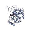
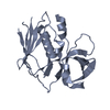
| Entry | Database: PDB / ID: 4drw | ||||||
|---|---|---|---|---|---|---|---|
| Title | Crystal Structure of the Ternary Complex between S100A10, an Annexin A2 N-terminal Peptide and an AHNAK Peptide | ||||||
 Components Components |
| ||||||
 Keywords Keywords | EXOCYTOSIS/PROTEIN BINDING / ATYPICAL EF-HAND / HETEROPENTAMERIC COMPLEX / MEMBRANE REPAIR / EXOCYTOSIS-PROTEIN BINDING complex | ||||||
| Function / homology |  Function and homology information Function and homology informationAnxA2-p11 complex / membrane raft assembly / positive regulation of receptor-mediated endocytosis involved in cholesterol transport / positive regulation of vacuole organization / phospholipase A2 inhibitor activity / positive regulation of low-density lipoprotein particle clearance / structural molecule activity conferring elasticity / negative regulation of low-density lipoprotein particle receptor catabolic process / positive regulation of plasma membrane repair / positive regulation of plasminogen activation ...AnxA2-p11 complex / membrane raft assembly / positive regulation of receptor-mediated endocytosis involved in cholesterol transport / positive regulation of vacuole organization / phospholipase A2 inhibitor activity / positive regulation of low-density lipoprotein particle clearance / structural molecule activity conferring elasticity / negative regulation of low-density lipoprotein particle receptor catabolic process / positive regulation of plasma membrane repair / positive regulation of plasminogen activation / PCSK9-AnxA2 complex / myelin sheath adaxonal region / osteoclast development / cadherin binding involved in cell-cell adhesion / positive regulation of vesicle fusion / cornified envelope / cell-cell contact zone / Schmidt-Lanterman incisure / vesicle budding from membrane / costamere / calcium-dependent phospholipid binding / negative regulation of receptor internalization / plasma membrane protein complex / Dissolution of Fibrin Clot / S100 protein binding / epithelial cell apoptotic process / collagen fibril organization / vesicle membrane / phosphatidylserine binding / regulation of RNA splicing / positive regulation of receptor recycling / positive regulation of exocytosis / basement membrane / positive regulation of focal adhesion assembly / positive regulation of GTPase activity / regulation of neurogenesis / Smooth Muscle Contraction / fibrinolysis / cytoskeletal protein binding / positive regulation of stress fiber assembly / positive regulation of substrate adhesion-dependent cell spreading / phosphatidylinositol-4,5-bisphosphate binding / lung development / lipid droplet / Gene and protein expression by JAK-STAT signaling after Interleukin-12 stimulation / T-tubule / Turbulent (oscillatory, disturbed) flow shear stress activates signaling by PIEZO1 and integrins in endothelial cells / cell-matrix adhesion / protein localization to plasma membrane / response to activity / Developmental Lineage of Pancreatic Ductal Cells / adherens junction / serine-type endopeptidase inhibitor activity / sarcolemma / mRNA transcription by RNA polymerase II / RNA polymerase II transcription regulator complex / nuclear matrix / calcium-dependent protein binding / azurophil granule lumen / melanosome / late endosome membrane / actin cytoskeleton / extracellular matrix / protease binding / angiogenesis / midbody / vesicle / basolateral plasma membrane / transmembrane transporter binding / early endosome / cell adhesion / endosome / cadherin binding / lysosomal membrane / focal adhesion / calcium ion binding / Neutrophil degranulation / cell surface / protein homodimerization activity / positive regulation of transcription by RNA polymerase II / : / RNA binding / extracellular exosome / extracellular region / membrane / identical protein binding / nucleus / plasma membrane / cytosol / cytoplasm Similarity search - Function | ||||||
| Biological species |  Homo sapiens (human) Homo sapiens (human) | ||||||
| Method |  X-RAY DIFFRACTION / SYNCHROTRON / MOLECULAR REPLACEMENT / Resolution: 3.5 Å X-RAY DIFFRACTION / SYNCHROTRON / MOLECULAR REPLACEMENT / Resolution: 3.5 Å | ||||||
 Authors Authors | Rezvanpour, A. / Lee, T.-W. / Junop, M.S. / Shaw, G.S. | ||||||
 Citation Citation | Journal: Structure / Year: 2012 Title: Structure of an asymmetric ternary protein complex provides insight for membrane interaction. Authors: Dempsey, B.R. / Rezvanpour, A. / Lee, T.W. / Barber, K.R. / Junop, M.S. / Shaw, G.S. | ||||||
| History |
|
- Structure visualization
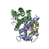
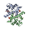
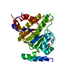
| Structure viewer | Molecule: MolmilJmol/JSmol |
|---|
- Downloads & links
-Download
| PDBx/mmCIF format | 4drw.cif.gz | 88.9 KB | Display | PDBx/mmCIF format |
|---|---|---|---|---|
| PDB format | pdb4drw.ent.gz | 70.3 KB | Display | PDB format |
| PDBx/mmJSON format | 4drw.json.gz | Tree view | PDBx/mmJSON format | |
| Others |  Other downloads Other downloads |
-Validation report
| Arichive directory | https://data.pdbj.org/pub/pdb/validation_reports/dr/4drwftp://data.pdbj.org/pub/pdb/validation_reports/dr/4drw | HTTPS FTP |
|---|
-Related structure data
| Related structure data |  1bt6S S: Starting model for refinement |
|---|---|
| Similar structure data |


-Links
 PDBj
PDBj


- Assembly
| Deposited unit | 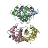
| ||||||||
|---|---|---|---|---|---|---|---|---|---|
| 1 | 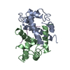
| ||||||||
| 2 | 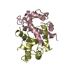
| ||||||||
| Unit cell |
|
-Components
| #1: Protein | Mass: 13765.778 Da / Num. of mol.: 4 Fragment: UNP P60903 residues 1-93 and UNP P07355 residues 2-16 Source method: isolated from a genetically manipulated source Source: (gene. exp.) Homo sapiens (human) / Gene: S100A10, ANX2LG, CAL1L, CLP11 / Production host:  #2: Protein/peptide | Mass: 2316.824 Da / Num. of mol.: 2 / Fragment: UNP Q09666 residues 5654-5673 / Source method: obtained synthetically / Details: CHEMICALLY SYNTHESIZED BASED ON HUMAN SEQUENCE / Source: (synth.) Homo sapiens (human) / References: UniProt: Q09666Has protein modification | Y | |
|---|
-Experimental details
-Experiment
| Experiment | Method: X-RAY DIFFRACTION / Number of used crystals: 2 |
|---|
- Sample preparation
| Crystal | Density Matthews: 2.07 Å3/Da / Density % sol: 40.49 % |
|---|---|
| Crystal grow | Temperature: 293 K / Method: vapor diffusion, hanging drop / pH: 6.5 Details: 100mM sodium chloride, 200mM magnesium chloride, 50mM sodium cacodylate, 20% PEG 1000, 0.15mM CYMAL-7, pH 6.5, VAPOR DIFFUSION, HANGING DROP, temperature 293K |
-Data collection
| Diffraction | Mean temperature: 100 K |
|---|---|
| Diffraction source | Source: SYNCHROTRON / Site: NSLS  / Beamline: X25 / Wavelength: 1.1 Å / Beamline: X25 / Wavelength: 1.1 Å |
| Detector | Type: ADSC QUANTUM 315 / Detector: CCD / Date: Oct 6, 2010 |
| Radiation | Monochromator: DOUBLE Si(111) CRYSTALS / Protocol: SINGLE WAVELENGTH / Monochromatic (M) / Laue (L): M / Scattering type: x-ray |
| Radiation wavelength | Wavelength: 1.1 Å / Relative weight: 1 |
| Reflection | Resolution: 3.26→43.34 Å / Num. all: 7410 / Num. obs: 7306 / % possible obs: 98.6 % / Observed criterion σ(F): 0 / Observed criterion σ(I): 0 |
| Reflection shell | Resolution: 3.26→3.32 Å / % possible all: 100 |
- Processing
| Software |
| ||||||||||||||||||||
|---|---|---|---|---|---|---|---|---|---|---|---|---|---|---|---|---|---|---|---|---|---|
| Refinement | Method to determine structure: MOLECULAR REPLACEMENT Starting model: PDB ENTRY 1BT6 Resolution: 3.5→43.34 Å / σ(F): 2 / Stereochemistry target values: Engh & Huber
| ||||||||||||||||||||
| Refinement step | Cycle: LAST / Resolution: 3.5→43.34 Å
| ||||||||||||||||||||
| Refine LS restraints |
|
