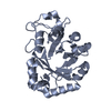
 Movie
Movie Controller


+ Open data

- Basic information
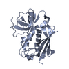
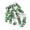
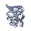
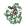
| Entry | Database: PDB / ID: 1cjm | ||||||
|---|---|---|---|---|---|---|---|
| Title | HUMAN SULT1A3 WITH SULFATE BOUND | ||||||
 Components Components | PROTEIN (ARYL SULFOTRANSFERASE) | ||||||
 Keywords Keywords | TRANSFERASE / SULT1A3 / HAST3 / SULFOTRANSFERASE / PAP / PAPS / DOPAMINE | ||||||
| Function / homology |  Function and homology information Function and homology information: / amine sulfotransferase activity / aryl sulfotransferase / flavonoid metabolic process / epinephrine metabolic process / aryl sulfotransferase activity / Cytosolic sulfonation of small molecules / sulfation / thyroid hormone metabolic process / 3'-phosphoadenosine 5'-phosphosulfate metabolic process ...: / amine sulfotransferase activity / aryl sulfotransferase / flavonoid metabolic process / epinephrine metabolic process / aryl sulfotransferase activity / Cytosolic sulfonation of small molecules / sulfation / thyroid hormone metabolic process / 3'-phosphoadenosine 5'-phosphosulfate metabolic process / sulfotransferase activity / ethanol catabolic process / norepinephrine metabolic process / sulfate binding / dopamine catabolic process / XBP1(S) activates chaperone genes / cellular response to dopamine / Paracetamol ADME / serotonin metabolic process / NMDA selective glutamate receptor signaling pathway / G protein-coupled dopamine receptor signaling pathway / steroid metabolic process / dopamine metabolic process / calcineurin-mediated signaling / ERK1 and ERK2 cascade / xenobiotic metabolic process / cytosol / cytoplasm Similarity search - Function | ||||||
| Biological species |  Homo sapiens (human) Homo sapiens (human) | ||||||
| Method |  X-RAY DIFFRACTION / SYNCHROTRON / MOLECULAR REPLACEMENT / Resolution: 2.4 Å X-RAY DIFFRACTION / SYNCHROTRON / MOLECULAR REPLACEMENT / Resolution: 2.4 Å | ||||||
 Authors Authors | Bidwell, L.M. / Mcmanus, M.E. / Gaedigk, A. / Kakuta, Y. / Negishi, M. / Pedersen, L. / Martin, J.L. | ||||||
 Citation Citation | Journal: J.Mol.Biol. / Year: 1999 Title: Crystal structure of human catecholamine sulfotransferase. Authors: Bidwell, L.M. / McManus, M.E. / Gaedigk, A. / Kakuta, Y. / Negishi, M. / Pedersen, L. / Martin, J.L. | ||||||
| History |
|
- Structure visualization
| Structure viewer | Molecule: MolmilJmol/JSmol |
|---|
- Downloads & links
-Download
| PDBx/mmCIF format | 1cjm.cif.gz | 59.2 KB | Display | PDBx/mmCIF format |
|---|---|---|---|---|
| PDB format | pdb1cjm.ent.gz | 41.7 KB | Display | PDB format |
| PDBx/mmJSON format | 1cjm.json.gz | Tree view | PDBx/mmJSON format | |
| Others |  Other downloads Other downloads |
-Validation report
| Arichive directory | https://data.pdbj.org/pub/pdb/validation_reports/cj/1cjmftp://data.pdbj.org/pub/pdb/validation_reports/cj/1cjm | HTTPS FTP |
|---|
-Related structure data
| Related structure data |  1aquS S: Starting model for refinement |
|---|---|
| Similar structure data |


-Links
 PDBj
PDBj
- Assembly
| Deposited unit | 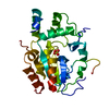
| ||||||||
|---|---|---|---|---|---|---|---|---|---|
| 1 |
| ||||||||
| Unit cell |
|
-Components
| #1: Protein | Mass: 34237.125 Da / Num. of mol.: 1 Source method: isolated from a genetically manipulated source Source: (gene. exp.) Homo sapiens (human) / Cellular location: CYTOPLASM / Organ: BRAIN (LIBRARY LAMBDA GT10) / Plasmid: PET20B / Cell line (production host): BL21(DE3) / Cellular location (production host): CYTOPLASM / Production host:  |
|---|---|
| #2: Chemical | ChemComp-SO4 /   Mass: 96.063 Da / Num. of mol.: 1 / Source method: obtained synthetically / Formula: SO4 Mass: 96.063 Da / Num. of mol.: 1 / Source method: obtained synthetically / Formula: SO4 |
| #3: Water | ChemComp-HOH /  Mass: 18.015 Da / Num. of mol.: 52 / Source method: isolated from a natural source / Formula: H2O Mass: 18.015 Da / Num. of mol.: 52 / Source method: isolated from a natural source / Formula: H2O |
-Experimental details
-Experiment
| Experiment | Method: X-RAY DIFFRACTION / Number of used crystals: 1 |
|---|
- Sample preparation
| Crystal | Density Matthews: 2.58 Å3/Da / Density % sol: 58 % | ||||||||||||||||||||||||||||||
|---|---|---|---|---|---|---|---|---|---|---|---|---|---|---|---|---|---|---|---|---|---|---|---|---|---|---|---|---|---|---|---|
| Crystal grow | pH: 7 Details: 0.5 M LITHIUM SULFATE AND 5-7% POLYETHYLENE GLYCOL 8000, pH 7.0 | ||||||||||||||||||||||||||||||
| Crystal | *PLUS | ||||||||||||||||||||||||||||||
| Crystal grow | *PLUS Temperature: 20 ℃ / pH: 6.8 / Method: vapor diffusion, hanging drop | ||||||||||||||||||||||||||||||
| Components of the solutions | *PLUS
|
-Data collection
| Diffraction | Mean temperature: 100 K |
|---|---|
| Diffraction source | Source: SYNCHROTRON / Site: SSRL  / Beamline: BL7-1 / Wavelength: 1.08 / Beamline: BL7-1 / Wavelength: 1.08 |
| Detector | Type: MARRESEARCH / Detector: IMAGE PLATE / Date: Mar 1, 1998 |
| Radiation | Protocol: SINGLE WAVELENGTH / Monochromatic (M) / Laue (L): M / Scattering type: x-ray |
| Radiation wavelength | Wavelength: 1.08 Å / Relative weight: 1 |
| Reflection | Resolution: 2.35→100 Å / Num. obs: 15080 / % possible obs: 97 % / Redundancy: 4.2 % / Biso Wilson estimate: 22.5 Å2 / Rsym value: 0.07 / Net I/σ(I): 14.3 |
| Reflection shell | Resolution: 2.35→2.43 Å / Mean I/σ(I) obs: 2.7 / Rsym value: 0.33 / % possible all: 87 |
| Reflection | *PLUS Observed criterion σ(I): 1 / Num. measured all: 63776 / Rmerge(I) obs: 0.07 |
| Reflection shell | *PLUS % possible obs: 86.8 % / Rmerge(I) obs: 0.33 |
- Processing
| Software |
| ||||||||||||||||||||||||||||||||||||||||||||||||||||||||||||||||||||||||||||||||
|---|---|---|---|---|---|---|---|---|---|---|---|---|---|---|---|---|---|---|---|---|---|---|---|---|---|---|---|---|---|---|---|---|---|---|---|---|---|---|---|---|---|---|---|---|---|---|---|---|---|---|---|---|---|---|---|---|---|---|---|---|---|---|---|---|---|---|---|---|---|---|---|---|---|---|---|---|---|---|---|---|---|
| Refinement | Method to determine structure: MOLECULAR REPLACEMENT Starting model: PDB ENTRY 1AQU Resolution: 2.4→100 Å / Rfactor Rfree error: 0.008 / Data cutoff high absF: 10000000 / Data cutoff low absF: 0.001 / Isotropic thermal model: RESTRAINED / Cross valid method: THROUGHOUT / σ(F): 0 / Details: BULK SOLVENT MODEL USED
| ||||||||||||||||||||||||||||||||||||||||||||||||||||||||||||||||||||||||||||||||
| Displacement parameters | Biso mean: 29 Å2
| ||||||||||||||||||||||||||||||||||||||||||||||||||||||||||||||||||||||||||||||||
| Refine analyze |
| ||||||||||||||||||||||||||||||||||||||||||||||||||||||||||||||||||||||||||||||||
| Refinement step | Cycle: LAST / Resolution: 2.4→100 Å
| ||||||||||||||||||||||||||||||||||||||||||||||||||||||||||||||||||||||||||||||||
| Refine LS restraints |
| ||||||||||||||||||||||||||||||||||||||||||||||||||||||||||||||||||||||||||||||||
| LS refinement shell | Resolution: 2.4→2.55 Å / Rfactor Rfree error: 0.021 / Total num. of bins used: 6
| ||||||||||||||||||||||||||||||||||||||||||||||||||||||||||||||||||||||||||||||||
| Xplor file |
| ||||||||||||||||||||||||||||||||||||||||||||||||||||||||||||||||||||||||||||||||
| Software | *PLUS Name: X-PLOR / Version: 3.851 / Classification: refinement | ||||||||||||||||||||||||||||||||||||||||||||||||||||||||||||||||||||||||||||||||
| Refinement | *PLUS σ(F): 0 / % reflection Rfree: 10.1 % / Rfactor obs: 0.276 | ||||||||||||||||||||||||||||||||||||||||||||||||||||||||||||||||||||||||||||||||
| Solvent computation | *PLUS | ||||||||||||||||||||||||||||||||||||||||||||||||||||||||||||||||||||||||||||||||
| Displacement parameters | *PLUS Biso mean: 29 Å2 | ||||||||||||||||||||||||||||||||||||||||||||||||||||||||||||||||||||||||||||||||
| Refine LS restraints | *PLUS
| ||||||||||||||||||||||||||||||||||||||||||||||||||||||||||||||||||||||||||||||||
| LS refinement shell | *PLUS Rfactor Rfree: 0.336 / % reflection Rfree: 11 % / Rfactor Rwork: 0.345 |
