- PDB-7h6g: THE 1.21 A CRYSTAL STRUCTURE OF HUMAN CATHEPSIN G IN COMPLEX WITH... -
+
Open data
ID or keywords:
Loading...
-
Basic information
Entry
Database: PDB / ID: 7h6g
Title
THE 1.21 A CRYSTAL STRUCTURE OF HUMAN CATHEPSIN G IN COMPLEX WITH N-[2-[6-fluoro-2-[(4-hydroxy-5-methyl-2-oxo-5-phenylfuran-3-yl)-phenylmethyl]-1H-indol-3-yl]ethyl]acetamide
Components
Cathepsin G
Keywords
HYDROLASE / HUMAN CATHEPSIN G / SERINE PROTEINASE / COMPLEX (SERINE PROTEASE-INHIBITOR) complex
Function / homology
Function and homology information
cathepsin G / biofilm matrix disassembly / neutrophil-mediated killing of gram-positive bacterium / purinergic nucleotide receptor signaling pathway / caspase binding / neutrophil activation / Suppression of apoptosis / Interleukin-1 processing / positive regulation of platelet aggregation / Antimicrobial peptides ...cathepsin G / biofilm matrix disassembly / neutrophil-mediated killing of gram-positive bacterium / purinergic nucleotide receptor signaling pathway / caspase binding / neutrophil activation / Suppression of apoptosis / Interleukin-1 processing / positive regulation of platelet aggregation / Antimicrobial peptides / negative regulation of T cell activation / Activation of Matrix Metalloproteinases / extracellular matrix disassembly / monocyte chemotaxis / defense response to fungus / Metabolism of Angiotensinogen to Angiotensins / Purinergic signaling in leishmaniasis infection / Degradation of the extracellular matrix / angiotensin maturation / secretory granule / serine-type peptidase activity / protein maturation / protein processing / platelet activation / positive regulation of immune response / Regulation of Insulin-like Growth Factor (IGF) transport and uptake by Insulin-like Growth Factor Binding Proteins (IGFBPs) / cytokine-mediated signaling pathway / cytoplasmic stress granule / azurophil granule lumen / peptidase activity / heparin binding / antibacterial humoral response / extracellular matrix / cellular response to lipopolysaccharide / defense response to Gram-negative bacterium / lysosome / defense response to Gram-positive bacterium / immune response / receptor ligand activity / serine-type endopeptidase activity / Neutrophil degranulation / proteolysis / : / extracellular exosome / extracellular region / membrane / nucleus / plasma membrane / cytosol Similarity search - Function
Serine proteases, trypsin family, histidine active site / Serine proteases, trypsin family, serine active site / Serine proteases, trypsin family, histidine active site. / Peptidase S1A, chymotrypsin family / Serine proteases, trypsin family, serine active site. / Serine proteases, trypsin domain profile. / Trypsin-like serine protease / Serine proteases, trypsin domain / Trypsin / Peptidase S1, PA clan, chymotrypsin-like fold / Peptidase S1, PA clan Similarity search - Domain/homology
Mass: 26801.787 Da / Num. of mol.: 2 Source method: isolated from a genetically manipulated source Source: (gene. exp.) Homo sapiens (human) / Gene: CTSG / Production host: Escherichia coli (E. coli) / References: UniProt: P08311, cathepsin G
Mass: 18.015 Da / Num. of mol.: 440 / Source method: isolated from a natural source / Formula: H2O
-
Details
Has protein modification
Y
-
Experimental details
-
Experiment
Experiment
Method: X-RAY DIFFRACTION / Number of used crystals: 1
-
Sample preparation
Crystal
Density Matthews: 2.15 Å3/Da / Density % sol: 42.72 %
Crystal grow
Temperature: 293 K / Method: microbatch / pH: 7.5 Details: 0.2 M LiSO4 (Salt), 25 %w/v PEG3350 (Precipitant), 0.1 M HEPES 7.5 pH (Buffer), Protein was at 17 mg/mL.
 Movie
Movie Controller

 Yorodumi
 Open data
 Basic information
 Components
 Keywords
 Function and homology information
 Homo sapiens (human)
 X-RAY DIFFRACTION /
  Authors
 Switzerland, 1items
  Citation
 Structure visualization
 Downloads & links
 Other downloads
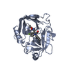


 PDBj
PDBj


 Assembly


 Mass: 65.409 Da / Num. of mol.: 4 / Source method: obtained synthetically / Formula: Zn
 Mass: 96.063 Da / Num. of mol.: 4 / Source method: obtained synthetically / Formula: SO4
 Mass: 209.240 Da / Num. of mol.: 1 / Source method: obtained synthetically / Formula: C8H19NO5 / Comment: pH buffer*YM
 Sample preparation
 Processing