- PDB-7h61: THE 1.76 A CRYSTAL STRUCTURE OF HUMAN CHYMASE IN COMPLEX WITH N-[... -
+
Open data
ID or keywords:
Loading...
-
Basic information
Entry
Database: PDB / ID: 7h61
Title
THE 1.76 A CRYSTAL STRUCTURE OF HUMAN CHYMASE IN COMPLEX WITH N-[2-[6-ethyl-2-[(2-hydroxy-5-oxo-3-phenylcyclopenten-1-yl)-phenylmethyl]-1H-indol-3-yl]ethyl]acetamide
Components
Chymase
Keywords
HYDROLASE / HUMAN CHYMASE / SERINE PROTEINASE
Function / homology
Function and homology information
chymase / basement membrane disassembly / cytokine precursor processing / peptide metabolic process / Activation of Matrix Metalloproteinases / midbrain development / extracellular matrix disassembly / Metabolism of Angiotensinogen to Angiotensins / angiotensin maturation / peptide binding ...chymase / basement membrane disassembly / cytokine precursor processing / peptide metabolic process / Activation of Matrix Metalloproteinases / midbrain development / extracellular matrix disassembly / Metabolism of Angiotensinogen to Angiotensins / angiotensin maturation / peptide binding / secretory granule / serine-type peptidase activity / protein catabolic process / cellular response to glucose stimulus / protein maturation / Signaling by SCF-KIT / positive regulation of angiogenesis / cytoplasmic ribonucleoprotein granule / extracellular matrix / regulation of inflammatory response / endopeptidase activity / serine-type endopeptidase activity / : / extracellular region / cytosol Similarity search - Function
Serine proteases, trypsin family, histidine active site / Serine proteases, trypsin family, serine active site / Serine proteases, trypsin family, histidine active site. / Peptidase S1A, chymotrypsin family / Serine proteases, trypsin family, serine active site. / Serine proteases, trypsin domain profile. / Trypsin-like serine protease / Serine proteases, trypsin domain / Trypsin / Peptidase S1, PA clan, chymotrypsin-like fold / Peptidase S1, PA clan Similarity search - Domain/homology
Mass: 18.015 Da / Num. of mol.: 375 / Source method: isolated from a natural source / Formula: H2O
Has protein modification
Y
-
Experimental details
-
Experiment
Experiment
Method: X-RAY DIFFRACTION / Number of used crystals: 1
-
Sample preparation
Crystal
Density Matthews: 2.75 Å3/Da / Density % sol: 55.32 %
Crystal grow
Temperature: 293 K / Method: microbatch / pH: 8.5 Details: Sample of human Chymase in 50 mM MES/NaOH pH 5.5, 150mM NaCl, 1mM TCEP, 10% Glycerol) at a concentration of 11mg/ml to 14mg/ml.Add [2-[(4-methylpyridin-2-yl)amino]-2-oxoethyl] 2- ...Details: Sample of human Chymase in 50 mM MES/NaOH pH 5.5, 150mM NaCl, 1mM TCEP, 10% Glycerol) at a concentration of 11mg/ml to 14mg/ml.Add [2-[(4-methylpyridin-2-yl)amino]-2-oxoethyl] 2-methylquinoline-4-carboxylate at 10x molar ratio. The compound helps to obtain crystals but is not visible in the structures. Add 0.5 mM ZnCl2. Add inhibitor, incubate for 16h on ice. Crystallize using microbatch setups with Al's oil (Hampton Research), with total drop size 1ul with 50% protein sample, using crystallization reagent of 0.1M Tris/HCl pH 8.5, 0.2M NaCl, 25% PEG 3350.
-
Data collection
Diffraction
Mean temperature: 100 K
Diffraction source
Source: ROTATING ANODE / Type: BRUKER AXS MICROSTAR / Wavelength: 1.5418 Å
Detector
Type: MAR scanner 345 mm plate / Detector: IMAGE PLATE / Date: Oct 25, 2005
Radiation
Protocol: SINGLE WAVELENGTH / Monochromatic (M) / Laue (L): M / Scattering type: x-ray
Radiation wavelength
Wavelength: 1.5418 Å / Relative weight: 1
Reflection
Resolution: 1.74→19.12 Å / Num. obs: 28058 / % possible obs: 99.8 % / Rmerge(I) obs: 0.045 / Rrim(I) all: 0.048 / Net I/σ(I): 31.67 / Num. measured all: 246195
Reflection shell
Diffraction-ID: 1
Resolution (Å)
% possible obs (%)
Rmerge(I) obs
Num. measured obs
Num. unique obs
Rrim(I) all
Net I/σ(I) obs
1.74-1.84
99.2
0.288
34152
4243
0.308
6.8
1.84-1.95
100
0.169
32466
3794
0.18
12.04
1.95-2.07
100
0.097
28327
3264
0.103
19.89
2.07-2.2
100
0.069
24354
2779
0.074
26.69
2.2-2.41
100
0.054
29539
3317
0.058
33.88
2.41-2.69
100
0.045
26870
2969
0.048
40.86
2.69-3.09
100
0.04
23888
2610
0.042
47.42
3.09-3.76
100
0.032
20641
2249
0.034
55.69
3.76-5.22
100
0.027
16266
1761
0.029
66.4
5.22-19.12
97.7
0.024
9692
1072
0.026
70.3
-
Processing
Software
Name
Version
Classification
REFMAC
5.8.0425
refinement
XSCALE
datascaling
XDS
datareduction
PHASER
phasing
Refinement
Method to determine structure: MOLECULAR REPLACEMENT / Resolution: 1.74→19.12 Å / Cor.coef. Fo:Fc: 0.965 / Cor.coef. Fo:Fc free: 0.954 / SU B: 1.71 / SU ML: 0.056 / Cross valid method: THROUGHOUT / ESU R: 0.1 / ESU R Free: 0.095 / Stereochemistry target values: MAXIMUM LIKELIHOOD / Details: HYDROGENS HAVE BEEN ADDED IN THE RIDING POSITIONS
Rfactor
Num. reflection
% reflection
Selection details
Rfree
0.18209
1410
5 %
RANDOM
Rwork
0.15546
-
-
-
obs
0.15678
26647
99.79 %
-
Solvent computation
Ion probe radii: 0.8 Å / Shrinkage radii: 0.8 Å / VDW probe radii: 1.2 Å / Solvent model: MASK
 Movie
Movie Controller

 Yorodumi
 Open data
 Basic information
 Components
 Keywords
 Function and homology information
 Homo sapiens (human)
 X-RAY DIFFRACTION /
  Authors
 Switzerland, 1items
  Citation
 Structure visualization
 Downloads & links
 Other downloads
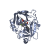


 PDBj
PDBj


 Assembly


 Mass: 65.409 Da / Num. of mol.: 1 / Source method: obtained synthetically / Formula: Zn
 Mass: 78.133 Da / Num. of mol.: 1 / Source method: obtained synthetically / Formula: C2H6OS / Comment: DMSO, precipitant*YM
 Mass: 18.015 Da / Num. of mol.: 375 / Source method: isolated from a natural source / Formula: H2O
 Sample preparation
 Processing