 Movie
Movie Controller

[English] 日本語
 Yorodumi
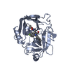
- PDB-7h63: THE 1.65 A CRYSTAL STRUCTURE OF HUMAN CHYMASE IN COMPLEX WITH 4-[... -
+ Open data

- Basic information
| Entry | Database: PDB / ID: 7h63 | ||||||
|---|---|---|---|---|---|---|---|
| Title | THE 1.65 A CRYSTAL STRUCTURE OF HUMAN CHYMASE IN COMPLEX WITH 4-[(5-fluoro-3-propan-2-yl-1H-indol-2-yl)-phenylmethyl]-3-hydroxy-2-propan-2-yl-1,2-dihydropyrrol-5-one (VINYLOGOUS ACID) | ||||||
 Components Components | Chymase | ||||||
 Keywords Keywords | HYDROLASE / HUMAN CHYMASE / SERINE PROTEINASE | ||||||
| Function / homology |  Function and homology information Function and homology informationchymase / basement membrane disassembly / cytokine precursor processing / peptide metabolic process / Activation of Matrix Metalloproteinases / midbrain development / extracellular matrix disassembly / Metabolism of Angiotensinogen to Angiotensins / angiotensin maturation / serine-type peptidase activity ...chymase / basement membrane disassembly / cytokine precursor processing / peptide metabolic process / Activation of Matrix Metalloproteinases / midbrain development / extracellular matrix disassembly / Metabolism of Angiotensinogen to Angiotensins / angiotensin maturation / serine-type peptidase activity / peptide binding / secretory granule / protein maturation / cellular response to glucose stimulus / protein catabolic process / Signaling by SCF-KIT / cytoplasmic ribonucleoprotein granule / positive regulation of angiogenesis / : / regulation of inflammatory response / endopeptidase activity / serine-type endopeptidase activity / extracellular space / extracellular region / cytosol Similarity search - Function | ||||||
| Biological species |  Homo sapiens (human) Homo sapiens (human) | ||||||
| Method |  X-RAY DIFFRACTION / MOLECULAR REPLACEMENT / Resolution: 1.65 Å X-RAY DIFFRACTION / MOLECULAR REPLACEMENT / Resolution: 1.65 Å | ||||||
 Authors Authors | Banner, D.W. / Benz, J.M. / Schlatter, D. / Hilpert, H. | ||||||
| Funding support |  Switzerland, 1items Switzerland, 1items
| ||||||
 Citation Citation | Journal: To be published Title: Crystal structures of human Chymase and Cathepsin G Authors: Markus, R. / Tosstorff, A. | ||||||
| History |
|
- Structure visualization
| Structure viewer | Molecule: MolmilJmol/JSmol |
|---|
- Downloads & links
-Download
| PDBx/mmCIF format | 7h63.cif.gz | 112.9 KB | Display | PDBx/mmCIF format |
|---|---|---|---|---|
| PDB format | pdb7h63.ent.gz | Display | PDB format | |
| PDBx/mmJSON format | 7h63.json.gz | Tree view | PDBx/mmJSON format | |
| Others |  Other downloads Other downloads |
-Validation report
| Summary document | 7h63_validation.pdf.gz | 804.4 KB | Display | wwPDB validaton report |
|---|---|---|---|---|
| Full document | 7h63_full_validation.pdf.gz | 804.9 KB | Display | |
| Data in XML | 7h63_validation.xml.gz | 16.1 KB | Display | |
| Data in CIF | 7h63_validation.cif.gz | 23.3 KB | Display | |
| Arichive directory | https://data.pdbj.org/pub/pdb/validation_reports/h6/7h63ftp://data.pdbj.org/pub/pdb/validation_reports/h6/7h63 | HTTPS FTP |
-Group deposition
| ID | G_1002292 (19 entries) |
|---|---|
| Title | A set of chymase crystal structures for D3R plus two CatG off-target structures |
| Type | undefined |
| Description | A set of chymase crystal structures for D3R plus two CatG off-target structures |


-Related structure data
| Similar structure data |
|---|
-Links
 PDBj
PDBj


- Assembly
| Deposited unit | 
| ||||||||
|---|---|---|---|---|---|---|---|---|---|
| 1 |
| ||||||||
| Unit cell |
|
-Components
| #1: Protein | Mass: 25032.910 Da / Num. of mol.: 1 / Mutation: C28S, F135K Source method: isolated from a genetically manipulated source Source: (gene. exp.) Homo sapiens (human) / Gene: CMA1, CYH, CYM / Production host:  |
|---|---|
| #2: Chemical | ChemComp-ZN /   Mass: 65.409 Da / Num. of mol.: 1 / Source method: obtained synthetically / Formula: Zn Mass: 65.409 Da / Num. of mol.: 1 / Source method: obtained synthetically / Formula: Zn |
| #3: Chemical | ChemComp-A1AOZ / ( Mass: 406.492 Da / Num. of mol.: 1 / Source method: obtained synthetically / Formula: C25H27FN2O2 / Feature type: SUBJECT OF INVESTIGATION |
| #4: Chemical | ChemComp-DMS /   Mass: 78.133 Da / Num. of mol.: 1 / Source method: obtained synthetically / Formula: C2H6OS / Comment: DMSO, precipitant*YM Mass: 78.133 Da / Num. of mol.: 1 / Source method: obtained synthetically / Formula: C2H6OS / Comment: DMSO, precipitant*YM |
| #5: Water | ChemComp-HOH /  Mass: 18.015 Da / Num. of mol.: 306 / Source method: isolated from a natural source / Formula: H2O Mass: 18.015 Da / Num. of mol.: 306 / Source method: isolated from a natural source / Formula: H2O |
| Has protein modification | Y |
-Experimental details
-Experiment
| Experiment | Method: X-RAY DIFFRACTION / Number of used crystals: 1 |
|---|
- Sample preparation
| Crystal | Density Matthews: 2.77 Å3/Da / Density % sol: 55.56 % |
|---|---|
| Crystal grow | Temperature: 293 K / Method: microbatch / pH: 8.5 Details: Sample of human Chymase in 50 mM MES/NaOH pH 5.5, 150mM NaCl, 1mM TCEP, 10% Glycerol) at a concentration of 11mg/ml to 14mg/ml.Add [2-[(4-methylpyridin-2-yl)amino]-2-oxoethyl] 2- ...Details: Sample of human Chymase in 50 mM MES/NaOH pH 5.5, 150mM NaCl, 1mM TCEP, 10% Glycerol) at a concentration of 11mg/ml to 14mg/ml.Add [2-[(4-methylpyridin-2-yl)amino]-2-oxoethyl] 2-methylquinoline-4-carboxylate at 10x molar ratio. The compound helps to obtain crystals but is not visible in the structures. Add 0.5 mM ZnCl2. Add inhibitor, incubate for 16h on ice. Crystallize using microbatch setups with Al's oil (Hampton Research), with total drop size 1ul with 50% protein sample, using crystallization reagent of 0.1M Tris/HCl pH 8.5, 0.2M NaCl, 25% PEG 3350. |
-Data collection
| Diffraction | Mean temperature: 100 K | ||||||||||||||||||||||||||||||||||||||||||||||||||||||||||||||||||||||
|---|---|---|---|---|---|---|---|---|---|---|---|---|---|---|---|---|---|---|---|---|---|---|---|---|---|---|---|---|---|---|---|---|---|---|---|---|---|---|---|---|---|---|---|---|---|---|---|---|---|---|---|---|---|---|---|---|---|---|---|---|---|---|---|---|---|---|---|---|---|---|---|
| Diffraction source | Source: ROTATING ANODE / Type: BRUKER AXS MICROSTAR / Wavelength: 1.5418 Å | ||||||||||||||||||||||||||||||||||||||||||||||||||||||||||||||||||||||
| Detector | Type: MAR scanner 345 mm plate / Detector: IMAGE PLATE / Date: Nov 25, 2005 | ||||||||||||||||||||||||||||||||||||||||||||||||||||||||||||||||||||||
| Radiation | Protocol: SINGLE WAVELENGTH / Monochromatic (M) / Laue (L): M / Scattering type: x-ray | ||||||||||||||||||||||||||||||||||||||||||||||||||||||||||||||||||||||
| Radiation wavelength | Wavelength: 1.5418 Å / Relative weight: 1 | ||||||||||||||||||||||||||||||||||||||||||||||||||||||||||||||||||||||
| Reflection | Resolution: 1.65→19.91 Å / Num. obs: 33005 / % possible obs: 99.6 % / Rmerge(I) obs: 0.062 / Rrim(I) all: 0.066 / Net I/σ(I): 22.18 / Num. measured all: 231352 | ||||||||||||||||||||||||||||||||||||||||||||||||||||||||||||||||||||||
| Reflection shell | Diffraction-ID: 1
|
- Processing
| Software |
| ||||||||||||||||||||||||||||||||||||||||||||||||||||||||||||||||||||||||||||||||||||||||||||||||||||||||||||||||||||||||||||||||||||||||||||||||||||||||||||||||||||||||||||||||||||||
|---|---|---|---|---|---|---|---|---|---|---|---|---|---|---|---|---|---|---|---|---|---|---|---|---|---|---|---|---|---|---|---|---|---|---|---|---|---|---|---|---|---|---|---|---|---|---|---|---|---|---|---|---|---|---|---|---|---|---|---|---|---|---|---|---|---|---|---|---|---|---|---|---|---|---|---|---|---|---|---|---|---|---|---|---|---|---|---|---|---|---|---|---|---|---|---|---|---|---|---|---|---|---|---|---|---|---|---|---|---|---|---|---|---|---|---|---|---|---|---|---|---|---|---|---|---|---|---|---|---|---|---|---|---|---|---|---|---|---|---|---|---|---|---|---|---|---|---|---|---|---|---|---|---|---|---|---|---|---|---|---|---|---|---|---|---|---|---|---|---|---|---|---|---|---|---|---|---|---|---|---|---|---|---|
| Refinement | Method to determine structure: MOLECULAR REPLACEMENT / Resolution: 1.65→19.91 Å / Cor.coef. Fo:Fc: 0.961 / Cor.coef. Fo:Fc free: 0.953 / SU B: 3.095 / SU ML: 0.056 / Cross valid method: THROUGHOUT / ESU R: 0.09 / ESU R Free: 0.086 / Stereochemistry target values: MAXIMUM LIKELIHOOD / Details: HYDROGENS HAVE BEEN ADDED IN THE RIDING POSITIONS
| ||||||||||||||||||||||||||||||||||||||||||||||||||||||||||||||||||||||||||||||||||||||||||||||||||||||||||||||||||||||||||||||||||||||||||||||||||||||||||||||||||||||||||||||||||||||
| Solvent computation | Ion probe radii: 0.8 Å / Shrinkage radii: 0.8 Å / VDW probe radii: 1.2 Å / Solvent model: MASK | ||||||||||||||||||||||||||||||||||||||||||||||||||||||||||||||||||||||||||||||||||||||||||||||||||||||||||||||||||||||||||||||||||||||||||||||||||||||||||||||||||||||||||||||||||||||
| Displacement parameters | Biso mean: 15.521 Å2
| ||||||||||||||||||||||||||||||||||||||||||||||||||||||||||||||||||||||||||||||||||||||||||||||||||||||||||||||||||||||||||||||||||||||||||||||||||||||||||||||||||||||||||||||||||||||
| Refinement step | Cycle: LAST / Resolution: 1.65→19.91 Å
| ||||||||||||||||||||||||||||||||||||||||||||||||||||||||||||||||||||||||||||||||||||||||||||||||||||||||||||||||||||||||||||||||||||||||||||||||||||||||||||||||||||||||||||||||||||||
| Refine LS restraints |
|