Movie
Movie Controller


+ Open data

- Basic information
| Entry | Database: PDB / ID: 7fd2 | |||||||||
|---|---|---|---|---|---|---|---|---|---|---|
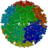
| Title | Cryo-EM structure of an alphavirus, Getah virus | |||||||||
 Components Components |
| |||||||||
 Keywords Keywords | VIRUS / mature / Infective | |||||||||
| Function / homology |  Function and homology information Function and homology informationT=4 icosahedral viral capsid / host cell cytoplasm / serine-type endopeptidase activity / fusion of virus membrane with host endosome membrane / symbiont entry into host cell / virion attachment to host cell / host cell plasma membrane / virion membrane / structural molecule activity / proteolysis / membrane Similarity search - Function | |||||||||
| Biological species |  Getah virus Getah virus | |||||||||
| Method | ELECTRON MICROSCOPY / single particle reconstruction / cryo EM / Resolution: 2.81 Å | |||||||||
 Authors Authors | Liu, Z. / Liu, C. / Wang, A. | |||||||||
| Funding support |  China, 2items China, 2items
| |||||||||
 Citation Citation | Journal: Cell Discov / Year: 2022 Title: Structure of infective Getah virus at 2.8 Å resolution determined by cryo-electron microscopy. Authors: Aojie Wang / Feng Zhou / Congcong Liu / Dongsheng Gao / Ruxi Qi / Yiheng Yin / Sheng Liu / Yuanzhu Gao / Lutang Fu / Yinhe Xia / Yawei Xu / Chuanqing Wang / Zheng Liu / Abstract: Getah virus (GETV), a member of the genus alphavirus, is a mosquito-borne pathogen that can cause pyrexia and reproductive losses in animals. Although antibodies to GETV have been found in over 10% ...Getah virus (GETV), a member of the genus alphavirus, is a mosquito-borne pathogen that can cause pyrexia and reproductive losses in animals. Although antibodies to GETV have been found in over 10% of healthy people, there are no reports of clinical symptoms associated with GETV. The biological and pathological properties of GETV are largely unknown and antiviral or vaccine treatments against GETV are still unavailable due to a lack of knowledge of the structure of the GETV virion. Here, we present the structure of infective GETV at a resolution of 2.8 Å with the atomic models of the capsid protein and the envelope glycoproteins E1 and E2. We have identified numerous glycosylation and S-acylation sites in E1 and E2. The surface-exposed glycans indicate a possible impact on viral immune evasion and host cell invasion. The S-acylation sites might be involved in stabilizing the transmembrane assembly of E1 and E2. In addition, a cholesterol and a phospholipid molecule are observed in a transmembrane hydrophobic pocket, together with two more cholesterols surrounding the pocket. The cholesterol and phospholipid stabilize the hydrophobic pocket in the viral envelope membrane. The structural information will assist structure-based antiviral and vaccine screening, design, and optimization. | |||||||||
| History |
|
- Structure visualization
| Structure viewer | Molecule: MolmilJmol/JSmol |
|---|
- Downloads & links
-Download
| PDBx/mmCIF format | 7fd2.cif.gz | 726.1 KB | Display | PDBx/mmCIF format |
|---|---|---|---|---|
| PDB format | pdb7fd2.ent.gz | 598.7 KB | Display | PDB format |
| PDBx/mmJSON format | 7fd2.json.gz | Tree view | PDBx/mmJSON format | |
| Others |  Other downloads Other downloads |
-Validation report
| Arichive directory | https://data.pdbj.org/pub/pdb/validation_reports/fd/7fd2ftp://data.pdbj.org/pub/pdb/validation_reports/fd/7fd2 | HTTPS FTP |
|---|
-Related structure data
| Related structure data | 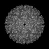 31533MC M: map data used to model this data C: citing same article ( |
|---|---|
| Similar structure data |


-Links
 PDBj
PDBj


- Assembly
| Deposited unit | 
|
|---|---|
| 1 | x 60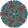
|
| 2 |
|
| 3 | x 5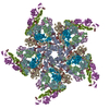
|
| 4 | x 6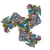
|
| 5 | 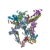
|
| Symmetry | Point symmetry: (Schoenflies symbol: I (icosahedral)) |
-Components
-Envelope glycoprotein ... , 2 types, 8 molecules AEIMBFJN
| #1: Protein | Mass: 47560.648 Da / Num. of mol.: 4 / Source method: isolated from a natural source / Source: (natural) Getah virus / References: UniProt: A0A1Z2R994, togavirin#2: Protein | Mass: 46147.492 Da / Num. of mol.: 4 / Source method: isolated from a natural source / Source: (natural) Getah virus / References: UniProt: A0A1Z2R994, togavirin |
|---|
-Protein , 1 types, 4 molecules CGKO
| #3: Protein | Mass: 30200.117 Da / Num. of mol.: 4 / Source method: isolated from a natural source / Source: (natural) Getah virus / References: UniProt: A0A1Z2R994, togavirin |
|---|
-Sugars , 4 types, 16 molecules 
| #4: Polysaccharide | alpha-D-mannopyranose-(1-6)-beta-D-mannopyranose-(1-4)-2-acetamido-2-deoxy-beta-D-glucopyranose-(1- ...alpha-D-mannopyranose-(1-6)-beta-D-mannopyranose-(1-4)-2-acetamido-2-deoxy-beta-D-glucopyranose-(1-4)-2-acetamido-2-deoxy-beta-D-glucopyranose Source method: isolated from a genetically manipulated source #5: Polysaccharide | 2-acetamido-2-deoxy-beta-D-glucopyranose-(1-4)-2-acetamido-2-deoxy-beta-D-glucopyranose Source method: isolated from a genetically manipulated source #6: Polysaccharide | beta-D-mannopyranose-(1-4)-2-acetamido-2-deoxy-beta-D-glucopyranose-(1-4)-2-acetamido-2-deoxy-beta- ...beta-D-mannopyranose-(1-4)-2-acetamido-2-deoxy-beta-D-glucopyranose-(1-4)-2-acetamido-2-deoxy-beta-D-glucopyranose Source method: isolated from a genetically manipulated source #7: Sugar | ChemComp-NAG /  Type: D-saccharide, beta linking / Mass: 221.208 Da / Num. of mol.: 4 / Source method: obtained synthetically / Formula: C8H15NO6 / Feature type: SUBJECT OF INVESTIGATION Type: D-saccharide, beta linking / Mass: 221.208 Da / Num. of mol.: 4 / Source method: obtained synthetically / Formula: C8H15NO6 / Feature type: SUBJECT OF INVESTIGATION |
|---|
-Non-polymers , 4 types, 36 molecules 


| #8: Chemical | ChemComp-PCW /  Mass: 787.121 Da / Num. of mol.: 4 / Source method: obtained synthetically / Formula: C44H85NO8P / Feature type: SUBJECT OF INVESTIGATION / Comment: DOPC, phospholipid*YM Mass: 787.121 Da / Num. of mol.: 4 / Source method: obtained synthetically / Formula: C44H85NO8P / Feature type: SUBJECT OF INVESTIGATION / Comment: DOPC, phospholipid*YM#9: Chemical | ChemComp-STE /  Mass: 284.477 Da / Num. of mol.: 8 / Source method: obtained synthetically / Formula: C18H36O2 / Feature type: SUBJECT OF INVESTIGATION Mass: 284.477 Da / Num. of mol.: 8 / Source method: obtained synthetically / Formula: C18H36O2 / Feature type: SUBJECT OF INVESTIGATION#10: Chemical | ChemComp-PLM /  Mass: 256.424 Da / Num. of mol.: 12 / Source method: obtained synthetically / Formula: C16H32O2 / Feature type: SUBJECT OF INVESTIGATION Mass: 256.424 Da / Num. of mol.: 12 / Source method: obtained synthetically / Formula: C16H32O2 / Feature type: SUBJECT OF INVESTIGATION#11: Chemical | ChemComp-CLR /  Mass: 386.654 Da / Num. of mol.: 12 / Source method: obtained synthetically / Formula: C27H46O / Feature type: SUBJECT OF INVESTIGATION Mass: 386.654 Da / Num. of mol.: 12 / Source method: obtained synthetically / Formula: C27H46O / Feature type: SUBJECT OF INVESTIGATION |
|---|
-Details
| Has ligand of interest | Y |
|---|---|
| Has protein modification | Y |
-Experimental details
-Experiment
| Experiment | Method: ELECTRON MICROSCOPY |
|---|---|
| EM experiment | Aggregation state: PARTICLE / 3D reconstruction method: single particle reconstruction |
- Sample preparation
| Component | Name: Getah virus / Type: VIRUS / Entity ID: #1-#3 / Source: NATURAL |
|---|---|
| Source (natural) | Organism: Getah virus / Strain: GETV-V1 |
| Details of virus | Empty: NO / Enveloped: YES / Isolate: STRAIN / Type: VIRION |
| Buffer solution | pH: 7.5 |
| Specimen | Embedding applied: NO / Shadowing applied: NO / Staining applied: NO / Vitrification applied: YES |
| Specimen support | Grid material: NICKEL/TITANIUM / Grid mesh size: 400 divisions/in. / Grid type: Quantifoil R2/2 |
| Vitrification | Cryogen name: ETHANE |
- Electron microscopy imaging
| Experimental equipment |  Model: Titan Krios / Image courtesy: FEI Company |
|---|---|
| Microscopy | Model: FEI TITAN KRIOS |
| Electron gun | Electron source:  FIELD EMISSION GUN / Accelerating voltage: 300 kV / Illumination mode: FLOOD BEAM FIELD EMISSION GUN / Accelerating voltage: 300 kV / Illumination mode: FLOOD BEAM |
| Electron lens | Mode: BRIGHT FIELD / Nominal magnification: 105000 X / Nominal defocus max: 1500 nm / Nominal defocus min: 800 nm / Calibrated defocus min: 1500 nm / Calibrated defocus max: 3000 nm / Cs: 2.7 mm / C2 aperture diameter: 70 µm / Alignment procedure: BASIC |
| Specimen holder | Cryogen: NITROGEN / Specimen holder model: FEI TITAN KRIOS AUTOGRID HOLDER |
| Image recording | Electron dose: 1.25 e/Å2 / Film or detector model: GATAN K3 BIOQUANTUM (6k x 4k) / Num. of grids imaged: 1 / Num. of real images: 16894 |
| Image scans | Width: 5760 / Height: 4092 |
- Processing
| Software | Name: PHENIX / Version: 1.19_4092: / Classification: refinement | ||||||||||||||||||||||||||||||||||||||||||||||||||
|---|---|---|---|---|---|---|---|---|---|---|---|---|---|---|---|---|---|---|---|---|---|---|---|---|---|---|---|---|---|---|---|---|---|---|---|---|---|---|---|---|---|---|---|---|---|---|---|---|---|---|---|
| EM software |
| ||||||||||||||||||||||||||||||||||||||||||||||||||
| CTF correction | Type: PHASE FLIPPING AND AMPLITUDE CORRECTION | ||||||||||||||||||||||||||||||||||||||||||||||||||
| Particle selection | Num. of particles selected: 171059 | ||||||||||||||||||||||||||||||||||||||||||||||||||
| 3D reconstruction | Resolution: 2.81 Å / Resolution method: FSC 0.143 CUT-OFF / Num. of particles: 2041957 / Num. of class averages: 1 / Symmetry type: POINT | ||||||||||||||||||||||||||||||||||||||||||||||||||
| Atomic model building | Protocol: RIGID BODY FIT / Space: REAL | ||||||||||||||||||||||||||||||||||||||||||||||||||
| Atomic model building | 3D fitting-ID: 1 / Accession code: 3J0C / Initial refinement model-ID: 1 / PDB-ID: 3J0C / Source name: PDB / Type: experimental model
| ||||||||||||||||||||||||||||||||||||||||||||||||||
| Refine LS restraints |
|