 Movie
Movie Controller

[English] 日本語
 Yorodumi
- PDB-7fbt: Crystal structure of chitinase (RmChi1) from Rhizomucor miehei (s... -
+ Open data

- Basic information
| Entry | Database: PDB / ID: 7fbt | |||||||||
|---|---|---|---|---|---|---|---|---|---|---|
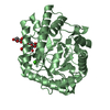
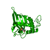
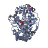
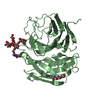
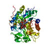
| Title | Crystal structure of chitinase (RmChi1) from Rhizomucor miehei (sp p32 2 1, MR) | |||||||||
 Components Components | Chitinase | |||||||||
 Keywords Keywords | HYDROLASE / Fungal chitinase / Rhizomucor miehei / P32 2 1 | |||||||||
| Function / homology |  Function and homology information Function and homology informationendochitinase activity / chitinase / chitin catabolic process / chitin binding / polysaccharide catabolic process / extracellular region / metal ion binding Similarity search - Function | |||||||||
| Biological species |  Rhizomucor miehei (fungus) Rhizomucor miehei (fungus) | |||||||||
| Method |  X-RAY DIFFRACTION / SYNCHROTRON / MOLECULAR REPLACEMENT / Resolution: 1.9 Å X-RAY DIFFRACTION / SYNCHROTRON / MOLECULAR REPLACEMENT / Resolution: 1.9 Å | |||||||||
 Authors Authors | Jiang, Z.Q. / Hu, S.Q. / Zhu, Q. / Liu, Y.C. / Ma, J.W. / Yan, Q.J. / Gao, Y.G. / Yang, S.Q. | |||||||||
 Citation Citation | Journal: Biochim Biophys Acta Proteins Proteom / Year: 2021 Title: Crystal structure of a chitinase (RmChiA) from the thermophilic fungus Rhizomucor miehei with a real active site tunnel. Authors: Jiang, Z. / Hu, S. / Ma, J. / Liu, Y. / Qiao, Z. / Yan, Q. / Gao, Y. / Yang, S. | |||||||||
| History |
|
- Structure visualization
| Structure viewer | Molecule: MolmilJmol/JSmol |
|---|
- Downloads & links
-Download
| PDBx/mmCIF format | 7fbt.cif.gz | 83.4 KB | Display | PDBx/mmCIF format |
|---|---|---|---|---|
| PDB format | pdb7fbt.ent.gz | 58.7 KB | Display | PDB format |
| PDBx/mmJSON format | 7fbt.json.gz | Tree view | PDBx/mmJSON format | |
| Others |  Other downloads Other downloads |
-Validation report
| Arichive directory | https://data.pdbj.org/pub/pdb/validation_reports/fb/7fbtftp://data.pdbj.org/pub/pdb/validation_reports/fb/7fbt | HTTPS FTP |
|---|
-Related structure data
| Related structure data |  5xwfC  5yuqC  1itxS C: citing same article ( S: Starting model for refinement |
|---|---|
| Similar structure data |


-Links
 PDBj
PDBj- Assembly
| Deposited unit | 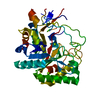
| ||||||||||
|---|---|---|---|---|---|---|---|---|---|---|---|
| 1 |
| ||||||||||
| Unit cell |
|
-Components
| #1: Protein | Mass: 41461.809 Da / Num. of mol.: 1 Source method: isolated from a genetically manipulated source Source: (gene. exp.) Rhizomucor miehei (fungus) / Production host:  | ||||||
|---|---|---|---|---|---|---|---|
| #2: Chemical |   Mass: 24.305 Da / Num. of mol.: 2 / Source method: obtained synthetically / Formula: Mg Mass: 24.305 Da / Num. of mol.: 2 / Source method: obtained synthetically / Formula: Mg#3: Water | ChemComp-HOH / |  Mass: 18.015 Da / Num. of mol.: 179 / Source method: isolated from a natural source / Formula: H2O Mass: 18.015 Da / Num. of mol.: 179 / Source method: isolated from a natural source / Formula: H2OHas ligand of interest | N | Has protein modification | Y | |
-Experimental details
-Experiment
| Experiment | Method: X-RAY DIFFRACTION / Number of used crystals: 1 |
|---|
- Sample preparation
| Crystal | Density Matthews: 1.81 Å3/Da / Density % sol: 31.89 % |
|---|---|
| Crystal grow | Temperature: 293.15 K / Method: vapor diffusion, sitting drop / pH: 8.5 Details: 200 mM MgCl2, 30 % (w/v) PEG 4000, 100 mM Tris-Cl pH 8.5 |
-Data collection
| Diffraction | Mean temperature: 100 K / Serial crystal experiment: N |
|---|---|
| Diffraction source | Source: SYNCHROTRON / Site: Photon Factory  / Beamline: BL-17A / Wavelength: 0.9643 Å / Beamline: BL-17A / Wavelength: 0.9643 Å |
| Detector | Type: ADSC QUANTUM 270 / Detector: CCD / Date: May 21, 2012 |
| Radiation | Protocol: SINGLE WAVELENGTH / Monochromatic (M) / Laue (L): M / Scattering type: x-ray |
| Radiation wavelength | Wavelength: 0.9643 Å / Relative weight: 1 |
| Reflection | Resolution: 1.9→33.29 Å / Num. obs: 23722 / % possible obs: 95.6 % / Redundancy: 27.82 % / Biso Wilson estimate: 32.88 Å2 / CC1/2: 1 / Rrim(I) all: 0.08 / Net I/σ(I): 30.94 |
| Reflection shell | Resolution: 1.9→2.02 Å / Redundancy: 11.66 % / Mean I/σ(I) obs: 3.46 / Num. unique obs: 3113 / CC1/2: 0.85 / Rrim(I) all: 0.75 / % possible all: 80.1 |
- Processing
| Software |
| ||||||||||||||||||||||||||||||||||||||||||||||||||||||||||||||||||||||||||||||||||||||||||||||||||||||||||||||||||||||||||||||
|---|---|---|---|---|---|---|---|---|---|---|---|---|---|---|---|---|---|---|---|---|---|---|---|---|---|---|---|---|---|---|---|---|---|---|---|---|---|---|---|---|---|---|---|---|---|---|---|---|---|---|---|---|---|---|---|---|---|---|---|---|---|---|---|---|---|---|---|---|---|---|---|---|---|---|---|---|---|---|---|---|---|---|---|---|---|---|---|---|---|---|---|---|---|---|---|---|---|---|---|---|---|---|---|---|---|---|---|---|---|---|---|---|---|---|---|---|---|---|---|---|---|---|---|---|---|---|---|
| Refinement | Method to determine structure: MOLECULAR REPLACEMENT Starting model: 1ITX Resolution: 1.9→33.29 Å / SU ML: 0.2762 / Cross valid method: FREE R-VALUE / σ(F): 1.37 / Phase error: 30.8514 Stereochemistry target values: GeoStd + Monomer Library + CDL v1.2
| ||||||||||||||||||||||||||||||||||||||||||||||||||||||||||||||||||||||||||||||||||||||||||||||||||||||||||||||||||||||||||||||
| Solvent computation | Shrinkage radii: 0.9 Å / VDW probe radii: 1.11 Å / Solvent model: FLAT BULK SOLVENT MODEL | ||||||||||||||||||||||||||||||||||||||||||||||||||||||||||||||||||||||||||||||||||||||||||||||||||||||||||||||||||||||||||||||
| Displacement parameters | Biso mean: 39.4 Å2 | ||||||||||||||||||||||||||||||||||||||||||||||||||||||||||||||||||||||||||||||||||||||||||||||||||||||||||||||||||||||||||||||
| Refinement step | Cycle: LAST / Resolution: 1.9→33.29 Å
| ||||||||||||||||||||||||||||||||||||||||||||||||||||||||||||||||||||||||||||||||||||||||||||||||||||||||||||||||||||||||||||||
| Refine LS restraints |
| ||||||||||||||||||||||||||||||||||||||||||||||||||||||||||||||||||||||||||||||||||||||||||||||||||||||||||||||||||||||||||||||
| LS refinement shell |
|
