 Movie
Movie Controller


+ Open data

- Basic information
| Entry | Database: PDB / ID: 1itx | ||||||
|---|---|---|---|---|---|---|---|
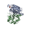
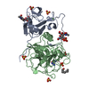
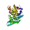
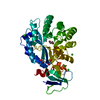
| Title | Catalytic Domain of Chitinase A1 from Bacillus circulans WL-12 | ||||||
 Components Components | Glycosyl Hydrolase | ||||||
 Keywords Keywords | HYDROLASE / Alpha-Beta (TIM) Barrel | ||||||
| Function / homology |  Function and homology information Function and homology informationendochitinase activity / chitinase / chitin catabolic process / chitin binding / polysaccharide catabolic process / carbohydrate binding / extracellular region Similarity search - Function | ||||||
| Biological species |  Bacillus circulans (bacteria) Bacillus circulans (bacteria) | ||||||
| Method |  X-RAY DIFFRACTION / SYNCHROTRON / MOLECULAR REPLACEMENT / Resolution: 1.1 Å X-RAY DIFFRACTION / SYNCHROTRON / MOLECULAR REPLACEMENT / Resolution: 1.1 Å | ||||||
 Authors Authors | Iwahori, F. / Matsumoto, T. / Watanabe, T. / Nonaka, T. | ||||||
 Citation Citation | Journal: PROC.JPN.ACAD.,SER.B / Year: 1999 Title: Three-dimensional structure of the catalytic domain of chitinase A1 from Bacillus circulans WL-12 at a very high resolution Authors: Matsumoto, T. / Nonaka, T. / Hashimoto, M. / Watanabe, T. / Mitsui, Y. #1: Journal: FEBS LETT. / Year: 2001Title: Trp122 and Trp134 on the surface of the catalytic domain are essential for crystalline chitin hydrolysis by Bacillus circulans chitinase A1 Authors: Watanabe, T. / Ishibashi, A. / Ariga, Y. / Hashimoto, M. / Nikaidou, N. / Sugiyama, J. / Matsumoto, T. / Nonaka, T. #2: Journal: PROTEIN PEPT.LETT. / Year: 1999Title: Crystallization and A Preliminary Crystallographic Analysis of the Catalytic Domain of Chitinase Al from Bacillus Circulans WL-12 Authors: Matsumoto, T. / Nonaka, T. / Katouda, H. / Hashimoto, M. / Watanabe, T. / Mitsui, Y. #3: Journal: J.BIOL.CHEM. / Year: 1993Title: Identification of glutamic acid 204 and aspartic acid 200 in chitinase A1 of Bacillus circulans WL-12 as essential residues for chitinase activity Authors: Watanabe, T. / Kobori, K. / Miyashita, K. / Fujii, T. / Sakai, H. / Uchida, M. / Tanaka, H. #4: Journal: J.BIOL.CHEM. / Year: 1990Title: Gene cloning of chitinase A1 from Bacillus circulans WL-12 revealed its evolutionary relationship to Serratia chitinase and to the type III homology units of fibronectin Authors: Watanabe, T. / Suzuki, K. / Oyanagi, W. / Ohnishi, K. / Tanaka, H. #5: Journal: J.BIOL.CHEM. / Year: 2001Title: Roles of the exposed aromatic residues in crystalline chitin hydrolysis by chitinase A from Serratia marcescens 2170 Authors: Uchiyama, T. / Katouno, f. / Nikaidou, N. / Nonaka, T. / Sugiyama, J. / Watanabe, T. | ||||||
| History |
|
- Structure visualization
| Structure viewer | Molecule: MolmilJmol/JSmol |
|---|
- Downloads & links
-Download
| PDBx/mmCIF format | 1itx.cif.gz | 216.5 KB | Display | PDBx/mmCIF format |
|---|---|---|---|---|
| PDB format | pdb1itx.ent.gz | 170.5 KB | Display | PDB format |
| PDBx/mmJSON format | 1itx.json.gz | Tree view | PDBx/mmJSON format | |
| Others |  Other downloads Other downloads |
-Validation report
| Arichive directory | https://data.pdbj.org/pub/pdb/validation_reports/it/1itxftp://data.pdbj.org/pub/pdb/validation_reports/it/1itx | HTTPS FTP |
|---|
-Related structure data
| Related structure data |  1ctnS S: Starting model for refinement |
|---|---|
| Similar structure data |


-Links
 PDBj
PDBj


- Assembly
| Deposited unit | 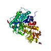
| ||||||||
|---|---|---|---|---|---|---|---|---|---|
| 1 |
| ||||||||
| Unit cell |
|
-Components
| #1: Protein | Mass: 45529.199 Da / Num. of mol.: 1 / Fragment: Catalytic Domain Source method: isolated from a genetically manipulated source Source: (gene. exp.) Bacillus circulans (bacteria) / Gene: CHIA1 / Plasmid: pKK223-3 / Production host: | ||||
|---|---|---|---|---|---|
| #2: Chemical | ChemComp-GOL /   Mass: 92.094 Da / Num. of mol.: 11 / Source method: obtained synthetically / Formula: C3H8O3 Mass: 92.094 Da / Num. of mol.: 11 / Source method: obtained synthetically / Formula: C3H8O3#3: Water | ChemComp-HOH / |  Mass: 18.015 Da / Num. of mol.: 770 / Source method: isolated from a natural source / Formula: H2O Mass: 18.015 Da / Num. of mol.: 770 / Source method: isolated from a natural source / Formula: H2OHas protein modification | Y | |
-Experimental details
-Experiment
| Experiment | Method: X-RAY DIFFRACTION / Number of used crystals: 1 |
|---|
- Sample preparation
| Crystal | Density Matthews: 1.97 Å3/Da / Density % sol: 37.21 % |
|---|---|
| Crystal grow | Temperature: 293 K / Method: vapor diffusion, hanging drop / pH: 5.3 Details: PEG 4000, potassium dihydrophosphate, pH 5.3, VAPOR DIFFUSION, HANGING DROP, temperature 293K |
-Data collection
| Diffraction | Mean temperature: 100 K |
|---|---|
| Diffraction source | Source: SYNCHROTRON / Site: SPring-8  / Beamline: BL41XU / Wavelength: 0.5 Å / Beamline: BL41XU / Wavelength: 0.5 Å |
| Detector | Type: MARRESEARCH / Detector: CCD / Date: May 18, 2001 |
| Radiation | Monochromator: undulator / Protocol: SINGLE WAVELENGTH / Monochromatic (M) / Laue (L): M / Scattering type: x-ray |
| Radiation wavelength | Wavelength: 0.5 Å / Relative weight: 1 |
| Reflection | Resolution: 1.1→36.793 Å / Num. all: 139823 / Num. obs: 139823 / % possible obs: 98.1 % / Observed criterion σ(F): 0 / Observed criterion σ(I): 0 / Redundancy: 1.9 % / Biso Wilson estimate: 5.943 Å2 / Rmerge(I) obs: 0.042 / Rsym value: 0.042 / Net I/σ(I): 10.1 |
| Reflection shell | Resolution: 1.1→1.13 Å / Redundancy: 1.9 % / Rmerge(I) obs: 0.248 / Mean I/σ(I) obs: 2.4 / Num. unique all: 10242 / Rsym value: 0.248 / % possible all: 97.2 |
- Processing
| Software |
| ||||||||||||||||||||||||||||||||||||||||||||||||||||||||||||||||||||||||||||||||||||||||||||||||||||||||||||||||||||||||||||||||||
|---|---|---|---|---|---|---|---|---|---|---|---|---|---|---|---|---|---|---|---|---|---|---|---|---|---|---|---|---|---|---|---|---|---|---|---|---|---|---|---|---|---|---|---|---|---|---|---|---|---|---|---|---|---|---|---|---|---|---|---|---|---|---|---|---|---|---|---|---|---|---|---|---|---|---|---|---|---|---|---|---|---|---|---|---|---|---|---|---|---|---|---|---|---|---|---|---|---|---|---|---|---|---|---|---|---|---|---|---|---|---|---|---|---|---|---|---|---|---|---|---|---|---|---|---|---|---|---|---|---|---|---|
| Refinement | Method to determine structure: MOLECULAR REPLACEMENT Starting model: PDB ENTRY 1CTN Resolution: 1.1→36.793 Å / Cor.coef. Fo:Fc: 0.984 / Cor.coef. Fo:Fc free: 0.979 / Isotropic thermal model: anisotropic / Cross valid method: THROUGHOUT / σ(F): 0 / σ(I): 0 / Stereochemistry target values: Engh & Huber Details: Some waters are listed as disorders coupled with some parts of the protein molecules.
| ||||||||||||||||||||||||||||||||||||||||||||||||||||||||||||||||||||||||||||||||||||||||||||||||||||||||||||||||||||||||||||||||||
| Solvent computation | Ion probe radii: 0.8 Å / Shrinkage radii: 0.8 Å / VDW probe radii: 1.4 Å / Solvent model: BABINET MODEL WITH MASK | ||||||||||||||||||||||||||||||||||||||||||||||||||||||||||||||||||||||||||||||||||||||||||||||||||||||||||||||||||||||||||||||||||
| Displacement parameters | Biso mean: 8.354 Å2 | ||||||||||||||||||||||||||||||||||||||||||||||||||||||||||||||||||||||||||||||||||||||||||||||||||||||||||||||||||||||||||||||||||
| Refinement step | Cycle: LAST / Resolution: 1.1→36.793 Å
| ||||||||||||||||||||||||||||||||||||||||||||||||||||||||||||||||||||||||||||||||||||||||||||||||||||||||||||||||||||||||||||||||||
| Refine LS restraints |
| ||||||||||||||||||||||||||||||||||||||||||||||||||||||||||||||||||||||||||||||||||||||||||||||||||||||||||||||||||||||||||||||||||
| LS refinement shell | Resolution: 1.1→1.129 Å / Total num. of bins used: 20
|
