 Movie
Movie Controller

[English] 日本語
 Yorodumi
- PDB-7f9n: Crystal structure of the variable region of Plasmodium RIFIN #4 (... -
+ Open data

- Basic information
| Entry | Database: PDB / ID: 7f9n | ||||||
|---|---|---|---|---|---|---|---|
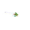
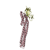
| Title | Crystal structure of the variable region of Plasmodium RIFIN #4 (PF3D7_1000500) in complex with LAIR1 | ||||||
 Components Components |
| ||||||
 Keywords Keywords | IMMUNE SYSTEM / malaria / Plasmodium falciparum / RIFIN / LAIR1 | ||||||
| Function / homology |  Function and homology information Function and homology informationimmune response-regulating signaling pathway / tertiary granule membrane / specific granule membrane / Immunoregulatory interactions between a Lymphoid and a non-Lymphoid cell / adaptive immune response / Neutrophil degranulation / plasma membrane Similarity search - Function | ||||||
| Biological species |   Homo sapiens (human) Homo sapiens (human) | ||||||
| Method |  X-RAY DIFFRACTION / SYNCHROTRON / MOLECULAR REPLACEMENT / Resolution: 3 Å X-RAY DIFFRACTION / SYNCHROTRON / MOLECULAR REPLACEMENT / Resolution: 3 Å | ||||||
 Authors Authors | Xie, Y. / Song, H. / Li, X. / Qi, J. / Gao, G.F. | ||||||
 Citation Citation | Journal: Cell Rep / Year: 2021 Title: Structural basis of malarial parasite RIFIN-mediated immune escape against LAIR1. Authors: Xie, Y. / Li, X. / Chai, Y. / Song, H. / Qi, J. / Gao, G.F. | ||||||
| History |
|
- Structure visualization
| Structure viewer | Molecule: MolmilJmol/JSmol |
|---|
- Downloads & links
-Download
| PDBx/mmCIF format | 7f9n.cif.gz | 133.1 KB | Display | PDBx/mmCIF format |
|---|---|---|---|---|
| PDB format | pdb7f9n.ent.gz | 84.6 KB | Display | PDB format |
| PDBx/mmJSON format | 7f9n.json.gz | Tree view | PDBx/mmJSON format | |
| Others |  Other downloads Other downloads |
-Validation report
| Arichive directory | https://data.pdbj.org/pub/pdb/validation_reports/f9/7f9nftp://data.pdbj.org/pub/pdb/validation_reports/f9/7f9n | HTTPS FTP |
|---|
-Related structure data
| Related structure data |  7f9kC 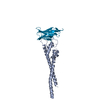 7f9lC 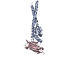 7f9mC  3kgrS S: Starting model for refinement C: citing same article ( |
|---|---|
| Similar structure data |


-Links
 PDBj
PDBj
- Assembly
| Deposited unit | 
| ||||||||||||
|---|---|---|---|---|---|---|---|---|---|---|---|---|---|
| 1 | 
| ||||||||||||
| 2 | 
| ||||||||||||
| Unit cell |
|
-Components
| #1: Protein | Mass: 18646.406 Da / Num. of mol.: 2 Source method: isolated from a genetically manipulated source Source: (gene. exp.) Strain: isolate 3D7 / Gene: PF3D7_1000500 / Production host:  #2: Protein | Mass: 12690.928 Da / Num. of mol.: 2 Source method: isolated from a genetically manipulated source Source: (gene. exp.) Homo sapiens (human) / Gene: LAIR1, CD305 / Production host: Has protein modification | Y | |
|---|
-Experimental details
-Experiment
| Experiment | Method: X-RAY DIFFRACTION / Number of used crystals: 1 |
|---|
- Sample preparation
| Crystal | Density Matthews: 3.67 Å3/Da / Density % sol: 66.47 % |
|---|---|
| Crystal grow | Temperature: 291 K / Method: vapor diffusion, sitting drop Details: 0.2 M di-ammonium hydrogen citrate, 20% w/v PEG 3,350 |
-Data collection
| Diffraction | Mean temperature: 100 K / Serial crystal experiment: N |
|---|---|
| Diffraction source | Source: SYNCHROTRON / Site: SSRF  / Beamline: BL17U / Wavelength: 0.97894 Å / Beamline: BL17U / Wavelength: 0.97894 Å |
| Detector | Type: SDMS / Detector: CCD / Date: Oct 2, 2018 |
| Radiation | Protocol: SINGLE WAVELENGTH / Monochromatic (M) / Laue (L): M / Scattering type: x-ray |
| Radiation wavelength | Wavelength: 0.97894 Å / Relative weight: 1 |
| Reflection | Resolution: 3→50 Å / Num. obs: 19913 / % possible obs: 100 % / Redundancy: 21 % / Biso Wilson estimate: 108.28 Å2 / CC1/2: 0.997 / Net I/σ(I): 21.7 |
| Reflection shell | Resolution: 3→3.11 Å / Num. unique obs: 19913 / CC1/2: 0.637 |
- Processing
| Software |
| ||||||||||||||||||||||||||||||||||||||||||||||||||||||||
|---|---|---|---|---|---|---|---|---|---|---|---|---|---|---|---|---|---|---|---|---|---|---|---|---|---|---|---|---|---|---|---|---|---|---|---|---|---|---|---|---|---|---|---|---|---|---|---|---|---|---|---|---|---|---|---|---|---|
| Refinement | Method to determine structure: MOLECULAR REPLACEMENT Starting model: 3KGR Resolution: 3→49.67 Å / SU ML: 0.5031 / Cross valid method: FREE R-VALUE / σ(F): 1.35 / Phase error: 40.436 / Stereochemistry target values: GeoStd + Monomer Library
| ||||||||||||||||||||||||||||||||||||||||||||||||||||||||
| Solvent computation | Shrinkage radii: 0.9 Å / VDW probe radii: 1.11 Å / Solvent model: FLAT BULK SOLVENT MODEL | ||||||||||||||||||||||||||||||||||||||||||||||||||||||||
| Displacement parameters | Biso mean: 116.33 Å2 | ||||||||||||||||||||||||||||||||||||||||||||||||||||||||
| Refinement step | Cycle: LAST / Resolution: 3→49.67 Å
| ||||||||||||||||||||||||||||||||||||||||||||||||||||||||
| Refine LS restraints |
| ||||||||||||||||||||||||||||||||||||||||||||||||||||||||
| LS refinement shell |
|
