| Entry | Database: PDB / ID: 5m04
|
|---|
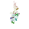
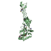
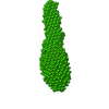
| Title | Structure of ObgE from Escherichia coli |
|---|
 Components Components | GTPase ObgE/CgtA |
|---|
 Keywords Keywords | HYDROLASE / GTPase / ObgE / CgtA |
|---|
| Function / homology |  Function and homology information Function and homology information
guanyl ribonucleotide binding / dormancy process / negative regulation of ribosome biogenesis / guanosine tetraphosphate binding / ribosomal large subunit binding / ribosome assembly / chromosome segregation / GDP binding / ribosomal large subunit assembly / Hydrolases; Acting on acid anhydrides; Acting on GTP to facilitate cellular and subcellular movement ...guanyl ribonucleotide binding / dormancy process / negative regulation of ribosome biogenesis / guanosine tetraphosphate binding / ribosomal large subunit binding / ribosome assembly / chromosome segregation / GDP binding / ribosomal large subunit assembly / Hydrolases; Acting on acid anhydrides; Acting on GTP to facilitate cellular and subcellular movement / rRNA binding / GTPase activity / GTP binding / magnesium ion binding / DNA binding / cytosolSimilarity search - Function Spo0b-associated Gtp-binding Protein; Chain: A, / GTP1/OBG domain / GTP1/OBG, conserved site / GTP1/OBG family signature. / GTP1/OBG domain / GTP-binding protein Obg/CgtA / GTP1/OBG domain superfamily / OBG-type GTPase / GTP1/OBG / Obg domain profile. ...Spo0b-associated Gtp-binding Protein; Chain: A, / GTP1/OBG domain / GTP1/OBG, conserved site / GTP1/OBG family signature. / GTP1/OBG domain / GTP-binding protein Obg/CgtA / GTP1/OBG domain superfamily / OBG-type GTPase / GTP1/OBG / Obg domain profile. / OBG-type guanine nucleotide-binding (G) domain / OBG-type guanine nucleotide-binding (G) domain profile. / 50S ribosome-binding GTPase / GTP binding domain / Distorted Sandwich / P-loop containing nucleotide triphosphate hydrolases / Rossmann fold / P-loop containing nucleoside triphosphate hydrolase / 3-Layer(aba) Sandwich / Mainly Beta / Alpha BetaSimilarity search - Domain/homology |
|---|
| Biological species |   Escherichia coli DH5[alpha] (bacteria) Escherichia coli DH5[alpha] (bacteria) |
|---|
| Method |  X-RAY DIFFRACTION / SYNCHROTRON / MOLECULAR REPLACEMENT / Resolution: 1.85 Å X-RAY DIFFRACTION / SYNCHROTRON / MOLECULAR REPLACEMENT / Resolution: 1.85 Å |
|---|
 Authors Authors | Gkekas, S. / Singh, R.K. / Versees, W. |
|---|
| Funding support |  Belgium, 2items Belgium, 2items | Organization | Grant number | Country |
|---|
| Fonds voor Wetenschappelijk Onderzoek (FWO) | G.0471.12N, G0B2515N | Belgium | | Institute for the Promotion of Innovation through Science and Technology in Flanders (IWT). | | Belgium |
|
|---|
 Citation Citation | Journal: J. Biol. Chem. / Year: 2017
Title: Structural and biochemical analysis of Escherichia coli ObgE, a central regulator of bacterial persistence.
Authors: Gkekas, S. / Singh, R.K. / Shkumatov, A.V. / Messens, J. / Fauvart, M. / Verstraeten, N. / Michiels, J. / Versees, W. |
|---|
| History | | Deposition | Oct 3, 2016 | Deposition site: PDBE / Processing site: PDBE |
|---|
| Revision 1.0 | Mar 1, 2017 | Provider: repository / Type: Initial release |
|---|
| Revision 1.1 | Mar 8, 2017 | Group: Database references |
|---|
| Revision 1.2 | Apr 19, 2017 | Group: Database references |
|---|
| Revision 1.3 | Jan 17, 2024 | Group: Data collection / Database references / Refinement description
Category: chem_comp_atom / chem_comp_bond ...chem_comp_atom / chem_comp_bond / database_2 / pdbx_initial_refinement_model
Item: _database_2.pdbx_DOI / _database_2.pdbx_database_accession |
|---|
|
|---|
 Movie
Movie Controller


 Open data
 Basic information
 Structure visualization
 Downloads & links
 Other downloads
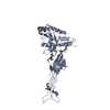


 PDBj
PDBj Assembly

 Type: RNA linking / Mass: 443.201 Da / Num. of mol.: 1 / Source method: obtained synthetically / Formula: C10H15N5O11P2 / Comment: GDP, energy-carrying molecule*YM
 Mass: 24.305 Da / Num. of mol.: 1 / Source method: obtained synthetically / Formula: Mg
 Mass: 18.015 Da / Num. of mol.: 169 / Source method: isolated from a natural source / Formula: H2O
 Sample preparation
 / Beamline: PROXIMA 2 / Wavelength: 1.07812 Å
 Processing