 Movie
Movie Controller

[English] 日本語
 Yorodumi
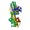
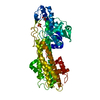
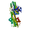
- PDB-7f8s: Pennisetum glaucum (Pearl millet) dehydroascorbate reductase (DHA... -
+ Open data

- Basic information
| Entry | Database: PDB / ID: 7f8s | ||||||||||||
|---|---|---|---|---|---|---|---|---|---|---|---|---|---|
| Title | Pennisetum glaucum (Pearl millet) dehydroascorbate reductase (DHAR) with catalytic cysteine (Cy20) in sulphenic and sulfinic acid forms. | ||||||||||||
 Components Components | (Dehydroascorbate reductase) x 2 | ||||||||||||
 Keywords Keywords | OXIDOREDUCTASE / Pennisetum glaucum / DHAR / Dehydroascorbate reductase / sulphenic acid / sulfinic acid / cysteine oxidation | ||||||||||||
| Function / homology |  Function and homology information Function and homology informationascorbate glutathione cycle / glutathione dehydrogenase (ascorbate) activity / glutathione dehydrogenase (ascorbate) / glutathione transferase activity Similarity search - Function | ||||||||||||
| Biological species |  Cenchrus americanus (pearl millet) Cenchrus americanus (pearl millet) | ||||||||||||
| Method |  X-RAY DIFFRACTION / MOLECULAR REPLACEMENT / Resolution: 2.63 Å X-RAY DIFFRACTION / MOLECULAR REPLACEMENT / Resolution: 2.63 Å | ||||||||||||
 Authors Authors | Das, B.K. / Kumar, A. / Sreeshma, N.S. / Arockiasamy, A. | ||||||||||||
| Funding support |  India, 3items India, 3items
| ||||||||||||
 Citation Citation | Journal: Biochem.Biophys.Res.Commun. / Year: 2021 Title: Comparative kinetic analysis of ascorbate (Vitamin-C) recycling dehydroascorbate reductases from plants and humans. Authors: Das, B.K. / Kumar, A. / Sreekumar, S.N. / Ponraj, K. / Gadave, K. / Kumar, S. / Murali Achary, V.M. / Ray, P. / Reddy, M.K. / Arockiasamy, A. #1: Journal: Biochem Biophys Res Commun / Year: 2016Title: Non-native ligands define the active site of Pennisetum glaucum (L.) R. Br dehydroascorbate reductase. Authors: Krishna Das, B. / Kumar, A. / Maindola, P. / Mahanty, S. / Jain, S.K. / Reddy, M.K. / Arockiasamy, A. | ||||||||||||
| History |
|
- Structure visualization
| Structure viewer | Molecule: MolmilJmol/JSmol |
|---|
- Downloads & links
-Download
| PDBx/mmCIF format | 7f8s.cif.gz | 98 KB | Display | PDBx/mmCIF format |
|---|---|---|---|---|
| PDB format | pdb7f8s.ent.gz | 72.7 KB | Display | PDB format |
| PDBx/mmJSON format | 7f8s.json.gz | Tree view | PDBx/mmJSON format | |
| Others |  Other downloads Other downloads |
-Validation report
| Arichive directory | https://data.pdbj.org/pub/pdb/validation_reports/f8/7f8sftp://data.pdbj.org/pub/pdb/validation_reports/f8/7f8s | HTTPS FTP |
|---|
-Related structure data
| Related structure data |  7f8rC  5evoS S: Starting model for refinement C: citing same article ( |
|---|---|
| Similar structure data |


-Links
 PDBj
PDBj

- Assembly
| Deposited unit | 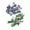
| ||||||||
|---|---|---|---|---|---|---|---|---|---|
| 1 | 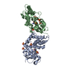
| ||||||||
| Unit cell |
|
-Components
| #1: Protein | Mass: 25631.301 Da / Num. of mol.: 1 Source method: isolated from a genetically manipulated source Source: (gene. exp.) Cenchrus americanus (pearl millet) / Production host:  References: UniProt: U5XYA0, glutathione dehydrogenase (ascorbate) | ||||||
|---|---|---|---|---|---|---|---|
| #2: Protein | Mass: 25647.301 Da / Num. of mol.: 1 Source method: isolated from a genetically manipulated source Source: (gene. exp.) Cenchrus americanus (pearl millet) / Production host: References: UniProt: U5XYA0, glutathione dehydrogenase (ascorbate) | ||||||
| #3: Chemical | ChemComp-SO4 /   Mass: 96.063 Da / Num. of mol.: 9 / Source method: obtained synthetically / Formula: SO4 Mass: 96.063 Da / Num. of mol.: 9 / Source method: obtained synthetically / Formula: SO4#4: Water | ChemComp-HOH / |  Mass: 18.015 Da / Num. of mol.: 86 / Source method: isolated from a natural source / Formula: H2O Mass: 18.015 Da / Num. of mol.: 86 / Source method: isolated from a natural source / Formula: H2OHas ligand of interest | Y | Has protein modification | Y | |
-Experimental details
-Experiment
| Experiment | Method: X-RAY DIFFRACTION / Number of used crystals: 1 |
|---|
- Sample preparation
| Crystal | Density Matthews: 2.53 Å3/Da / Density % sol: 51.44 % / Description: Bipyramidal crystals |
|---|---|
| Crystal grow | Temperature: 293.15 K / Method: vapor diffusion, sitting drop / pH: 4.6 / Details: 0.1M Sodium acetate pH 4.6, 2.0M Ammonium sulphate |
-Data collection
| Diffraction | Mean temperature: 100 K / Serial crystal experiment: N | |||||||||||||||||||||||||||||||||||||||||||||||||||||||||||||||||||||||||||||||||||||||||||||||||||||||||||||||||||||||||||||||||||||||||||||||||||||||||||||||||||||||||||||||||||||||||||||
|---|---|---|---|---|---|---|---|---|---|---|---|---|---|---|---|---|---|---|---|---|---|---|---|---|---|---|---|---|---|---|---|---|---|---|---|---|---|---|---|---|---|---|---|---|---|---|---|---|---|---|---|---|---|---|---|---|---|---|---|---|---|---|---|---|---|---|---|---|---|---|---|---|---|---|---|---|---|---|---|---|---|---|---|---|---|---|---|---|---|---|---|---|---|---|---|---|---|---|---|---|---|---|---|---|---|---|---|---|---|---|---|---|---|---|---|---|---|---|---|---|---|---|---|---|---|---|---|---|---|---|---|---|---|---|---|---|---|---|---|---|---|---|---|---|---|---|---|---|---|---|---|---|---|---|---|---|---|---|---|---|---|---|---|---|---|---|---|---|---|---|---|---|---|---|---|---|---|---|---|---|---|---|---|---|---|---|---|---|---|---|
| Diffraction source | Source: ROTATING ANODE / Type: RIGAKU MICROMAX-007 / Wavelength: 1.5418 Å | |||||||||||||||||||||||||||||||||||||||||||||||||||||||||||||||||||||||||||||||||||||||||||||||||||||||||||||||||||||||||||||||||||||||||||||||||||||||||||||||||||||||||||||||||||||||||||||
| Detector | Type: MAR scanner 345 mm plate / Detector: IMAGE PLATE / Date: Aug 1, 2013 | |||||||||||||||||||||||||||||||||||||||||||||||||||||||||||||||||||||||||||||||||||||||||||||||||||||||||||||||||||||||||||||||||||||||||||||||||||||||||||||||||||||||||||||||||||||||||||||
| Radiation | Protocol: SINGLE WAVELENGTH / Monochromatic (M) / Laue (L): M / Scattering type: x-ray | |||||||||||||||||||||||||||||||||||||||||||||||||||||||||||||||||||||||||||||||||||||||||||||||||||||||||||||||||||||||||||||||||||||||||||||||||||||||||||||||||||||||||||||||||||||||||||||
| Radiation wavelength | Wavelength: 1.5418 Å / Relative weight: 1 | |||||||||||||||||||||||||||||||||||||||||||||||||||||||||||||||||||||||||||||||||||||||||||||||||||||||||||||||||||||||||||||||||||||||||||||||||||||||||||||||||||||||||||||||||||||||||||||
| Reflection | Resolution: 2.63→50 Å / Num. obs: 16093 / % possible obs: 99.1 % / Redundancy: 13.6 % / Biso Wilson estimate: 50.44 Å2 / Rmerge(I) obs: 0.151 / Rpim(I) all: 0.042 / Rrim(I) all: 0.157 / Χ2: 0.675 / Net I/σ(I): 4.3 / Num. measured all: 219205 | |||||||||||||||||||||||||||||||||||||||||||||||||||||||||||||||||||||||||||||||||||||||||||||||||||||||||||||||||||||||||||||||||||||||||||||||||||||||||||||||||||||||||||||||||||||||||||||
| Reflection shell | Diffraction-ID: 1
|
- Processing
| Software |
| ||||||||||||||||||||||||||||||||||||||||||||||||||||||||||||||||||||||||||||||||||||||||||||||||||||||||||||||||||
|---|---|---|---|---|---|---|---|---|---|---|---|---|---|---|---|---|---|---|---|---|---|---|---|---|---|---|---|---|---|---|---|---|---|---|---|---|---|---|---|---|---|---|---|---|---|---|---|---|---|---|---|---|---|---|---|---|---|---|---|---|---|---|---|---|---|---|---|---|---|---|---|---|---|---|---|---|---|---|---|---|---|---|---|---|---|---|---|---|---|---|---|---|---|---|---|---|---|---|---|---|---|---|---|---|---|---|---|---|---|---|---|---|---|---|---|
| Refinement | Method to determine structure: MOLECULAR REPLACEMENT Starting model: 5EVO Resolution: 2.63→48.78 Å / Cor.coef. Fo:Fc: 0.926 / Cor.coef. Fo:Fc free: 0.894 / SU R Cruickshank DPI: 0.62 / Cross valid method: THROUGHOUT / σ(F): 0 / SU R Blow DPI: 0.713 / SU Rfree Blow DPI: 0.307 / SU Rfree Cruickshank DPI: 0.306
| ||||||||||||||||||||||||||||||||||||||||||||||||||||||||||||||||||||||||||||||||||||||||||||||||||||||||||||||||||
| Displacement parameters | Biso mean: 35.78 Å2
| ||||||||||||||||||||||||||||||||||||||||||||||||||||||||||||||||||||||||||||||||||||||||||||||||||||||||||||||||||
| Refine analyze | Luzzati coordinate error obs: 0.33 Å | ||||||||||||||||||||||||||||||||||||||||||||||||||||||||||||||||||||||||||||||||||||||||||||||||||||||||||||||||||
| Refinement step | Cycle: 1 / Resolution: 2.63→48.78 Å
| ||||||||||||||||||||||||||||||||||||||||||||||||||||||||||||||||||||||||||||||||||||||||||||||||||||||||||||||||||
| Refine LS restraints |
| ||||||||||||||||||||||||||||||||||||||||||||||||||||||||||||||||||||||||||||||||||||||||||||||||||||||||||||||||||
| LS refinement shell | Resolution: 2.63→2.66 Å / Rfactor Rfree error: 0
|
