 Movie
Movie Controller

[English] 日本語
 Yorodumi
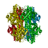
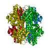
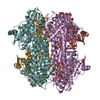
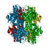
- PDB-1gk3: Histidine Ammonia-Lyase (HAL) Mutant D145A from Pseudomonas putida -
+ Open data

- Basic information
| Entry | Database: PDB / ID: 1gk3 | ||||||
|---|---|---|---|---|---|---|---|
| Title | Histidine Ammonia-Lyase (HAL) Mutant D145A from Pseudomonas putida | ||||||
 Components Components | HISTIDINE AMMONIA-LYASE | ||||||
 Keywords Keywords | LYASE / AMMONIA-LYASE / HISTIDINE DEGRADATION | ||||||
| Function / homology |  Function and homology information Function and homology informationhistidine ammonia-lyase / histidine ammonia-lyase activity / : / : / cytoplasm Similarity search - Function | ||||||
| Biological species |  PSEUDOMONAS PUTIDA (bacteria) PSEUDOMONAS PUTIDA (bacteria) | ||||||
| Method |  X-RAY DIFFRACTION / OTHER / Resolution: 2.25 Å X-RAY DIFFRACTION / OTHER / Resolution: 2.25 Å | ||||||
 Authors Authors | Baedeker, M. / Schulz, G.E. | ||||||
 Citation Citation | Journal: Structure / Year: 2002 Title: Autocatalytic Peptide Cyclization During Chain Folding of Histidine Ammonia-Lyase. Authors: Baedeker, M. / Schulz, G.E. #1: Journal: Biochemistry / Year: 1999Title: Crystal Structure of Histidine Ammonia-Lyase Revealing a Novel Polypeptide Modification as the Catalytic Electrophile Authors: Schwede, T.F. / Retey, J. / Schulz, G.E. #2: Journal: Protein Eng. / Year: 1999 Title: Homogenization and Crystallization of Histidine Ammonia-Lyase by Exchange of a Surface Cysteine Residue Authors: Schwede, T.F. / Baedeker, M. / Langer, M. / Retey, J. / Schulz, G.E. | ||||||
| History |
|
- Structure visualization
| Structure viewer | Molecule: MolmilJmol/JSmol |
|---|
- Downloads & links
-Download
| PDBx/mmCIF format | 1gk3.cif.gz | 109.9 KB | Display | PDBx/mmCIF format |
|---|---|---|---|---|
| PDB format | pdb1gk3.ent.gz | 84.8 KB | Display | PDB format |
| PDBx/mmJSON format | 1gk3.json.gz | Tree view | PDBx/mmJSON format | |
| Others |  Other downloads Other downloads |
-Validation report
| Arichive directory | https://data.pdbj.org/pub/pdb/validation_reports/gk/1gk3ftp://data.pdbj.org/pub/pdb/validation_reports/gk/1gk3 | HTTPS FTP |
|---|
-Related structure data


-Links
 PDBj
PDBj

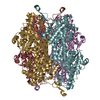
- Assembly
| Deposited unit | 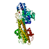
| |||||||||||||||
|---|---|---|---|---|---|---|---|---|---|---|---|---|---|---|---|---|
| 1 | 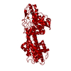
| |||||||||||||||
| Unit cell |
| |||||||||||||||
| Components on special symmetry positions |
|
-Components
| #1: Protein | Mass: 53611.246 Da / Num. of mol.: 1 / Mutation: YES Source method: isolated from a genetically manipulated source Details: THIS MUTANT DOES NOT CONTAIN A 4-METHYLIDENE-IMIDAZOLE-5-ONE GROUP. Source: (gene. exp.) PSEUDOMONAS PUTIDA (bacteria) / Plasmid: PT7-7H / Production host: |
|---|---|
| #2: Chemical | ChemComp-SO4 /   Mass: 96.063 Da / Num. of mol.: 1 / Source method: obtained synthetically / Formula: SO4 Mass: 96.063 Da / Num. of mol.: 1 / Source method: obtained synthetically / Formula: SO4 |
| #3: Chemical | ChemComp-GOL /   Mass: 92.094 Da / Num. of mol.: 1 / Source method: obtained synthetically / Formula: C3H8O3 Mass: 92.094 Da / Num. of mol.: 1 / Source method: obtained synthetically / Formula: C3H8O3 |
| #4: Water | ChemComp-HOH /  Mass: 18.015 Da / Num. of mol.: 184 / Source method: isolated from a natural source / Formula: H2O Mass: 18.015 Da / Num. of mol.: 184 / Source method: isolated from a natural source / Formula: H2O |
| Compound details | CHAIN A ENGINEERED MUTATION CYS273ALA, ASP145ALA MUTANT D145A IS UNABLE TO FORM THE CATALYTICALLY ...CHAIN A ENGINEERED |
-Experimental details
-Experiment
| Experiment | Method: X-RAY DIFFRACTION / Number of used crystals: 1 |
|---|
- Sample preparation
| Crystal | Density Matthews: 2.8 Å3/Da / Density % sol: 58.06 % | ||||||||||||||||||||||||||||||||||||
|---|---|---|---|---|---|---|---|---|---|---|---|---|---|---|---|---|---|---|---|---|---|---|---|---|---|---|---|---|---|---|---|---|---|---|---|---|---|
| Crystal grow | pH: 8.1 Details: CRYSTALLIZED FROM 2.0 M (NH4)2SO4, 1 % GLYCEROL, 2 % PEG 400, 0.1 M HEPES AT PH 8.1. 20 % (V/V) GLYCEROL WERE USED AS CRYOPROTECTANT | ||||||||||||||||||||||||||||||||||||
| Crystal grow | *PLUS pH: 3.85 / Method: vapor diffusion, hanging drop / Details: Schwede, T.F., (1999) Protein Eng., 12, 151. | ||||||||||||||||||||||||||||||||||||
| Components of the solutions | *PLUS
|
-Data collection
| Diffraction | Mean temperature: 100 K |
|---|---|
| Diffraction source | Source: ROTATING ANODE / Type: RIGAKU RUB200 / Wavelength: 1.5418 |
| Detector | Type: MARRESEARCH / Detector: IMAGE PLATE |
| Radiation | Monochromator: GRAPHITE CRYSTAL / Protocol: SINGLE WAVELENGTH / Monochromatic (M) / Laue (L): M / Scattering type: x-ray |
| Radiation wavelength | Wavelength: 1.5418 Å / Relative weight: 1 |
| Reflection | Resolution: 2.25→21 Å / Num. obs: 26022 / % possible obs: 91 % / Redundancy: 2.3 % / Rmerge(I) obs: 0.126 / Net I/σ(I): 2.4 |
| Reflection shell | *PLUS % possible obs: 95 % / Redundancy: 2.2 % / Rmerge(I) obs: 0.29 / Mean I/σ(I) obs: 1.4 |
- Processing
| Software |
| |||||||||||||||||||||||||||||||||
|---|---|---|---|---|---|---|---|---|---|---|---|---|---|---|---|---|---|---|---|---|---|---|---|---|---|---|---|---|---|---|---|---|---|---|
| Refinement | Method to determine structure: OTHER / Resolution: 2.25→21 Å / Cross valid method: THROUGHOUT / σ(F): 0 Details: RESIDUES 272-275 AND 363-374 NOT VISIBLE IN ELECTRON DENSITY
| |||||||||||||||||||||||||||||||||
| Refinement step | Cycle: LAST / Resolution: 2.25→21 Å
| |||||||||||||||||||||||||||||||||
| Refine LS restraints |
| |||||||||||||||||||||||||||||||||
| Software | *PLUS Name: SHELX / Classification: refinement | |||||||||||||||||||||||||||||||||
| Refinement | *PLUS Rfactor Rfree: 0.28 / Rfactor Rwork: 0.221 | |||||||||||||||||||||||||||||||||
| Solvent computation | *PLUS | |||||||||||||||||||||||||||||||||
| Displacement parameters | *PLUS |
