 Movie
Movie Controller


+ Open data

- Basic information
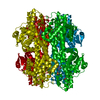
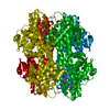
| Entry | Database: PDB / ID: 1b8f | ||||||||||||
|---|---|---|---|---|---|---|---|---|---|---|---|---|---|
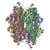
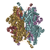
| Title | Histidine ammonia-lyase (HAL) from Pseudomonas putida | ||||||||||||
 Components Components | Histidine ammonia-lyase | ||||||||||||
 Keywords Keywords | LYASE / AMMONIA-LYASE / HISTIDINE DEGRADATION | ||||||||||||
| Function / homology |  Function and homology information Function and homology informationhistidine ammonia-lyase / histidine ammonia-lyase activity / : / : / cytoplasm Similarity search - Function | ||||||||||||
| Biological species |  Pseudomonas putida (bacteria) Pseudomonas putida (bacteria) | ||||||||||||
| Method |  X-RAY DIFFRACTION / MOLECULAR REPLACEMENT / Resolution: 2.1 Å X-RAY DIFFRACTION / MOLECULAR REPLACEMENT / Resolution: 2.1 Å | ||||||||||||
 Authors Authors | Schwede, T.F. / Schulz, G.E. | ||||||||||||
 Citation Citation | Journal: Biochemistry / Year: 1999 Title: Crystal structure of histidine ammonia-lyase revealing a novel polypeptide modification as the catalytic electrophile. Authors: Schwede, T.F. / Retey, J. / Schulz, G.E. #1: Journal: Protein Eng. / Year: 1999 Title: Homogenization and crystallization of histidine ammonia-lyase by exchange of a surface cysteine residue. Authors: Schwede, T.F. / Badeker, M. / Langer, M. / Retey, J. / Schulz, G.E. | ||||||||||||
| History |
|
- Structure visualization
| Structure viewer | Molecule: MolmilJmol/JSmol |
|---|
- Downloads & links
-Download
| PDBx/mmCIF format | 1b8f.cif.gz | 110.8 KB | Display | PDBx/mmCIF format |
|---|---|---|---|---|
| PDB format | pdb1b8f.ent.gz | 83.7 KB | Display | PDB format |
| PDBx/mmJSON format | 1b8f.json.gz | Tree view | PDBx/mmJSON format | |
| Others |  Other downloads Other downloads |
-Validation report
| Arichive directory | https://data.pdbj.org/pub/pdb/validation_reports/b8/1b8fftp://data.pdbj.org/pub/pdb/validation_reports/b8/1b8f | HTTPS FTP |
|---|
-Related structure data
| Similar structure data |
|---|


-Links
 PDBj
PDBj

- Assembly
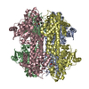
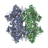
| Deposited unit | 
| ||||||||
|---|---|---|---|---|---|---|---|---|---|
| 1 | 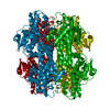
| ||||||||
| Unit cell |
|
-Components
| #1: Protein | Mass: 53619.223 Da / Num. of mol.: 1 / Mutation: C273A Source method: isolated from a genetically manipulated source Source: (gene. exp.) Pseudomonas putida (bacteria) / Cellular location: CYTOPLASM / Gene: hutH / Plasmid: PT7-7H / Cellular location (production host): CYTOPLASM / Production host: |
|---|---|
| #2: Chemical | ChemComp-SO4 /   Mass: 96.063 Da / Num. of mol.: 1 / Source method: obtained synthetically / Formula: SO4 Mass: 96.063 Da / Num. of mol.: 1 / Source method: obtained synthetically / Formula: SO4 |
| #3: Chemical | ChemComp-GOL /   Mass: 92.094 Da / Num. of mol.: 1 / Source method: obtained synthetically / Formula: C3H8O3 Mass: 92.094 Da / Num. of mol.: 1 / Source method: obtained synthetically / Formula: C3H8O3 |
| #4: Water | ChemComp-HOH /  Mass: 18.015 Da / Num. of mol.: 210 / Source method: isolated from a natural source / Formula: H2O Mass: 18.015 Da / Num. of mol.: 210 / Source method: isolated from a natural source / Formula: H2O |
| Has protein modification | Y |
-Experimental details
-Experiment
| Experiment | Method: X-RAY DIFFRACTION / Number of used crystals: 1 |
|---|
- Sample preparation
| Crystal | Density Matthews: 2.8 Å3/Da / Density % sol: 56 % | ||||||||||||||||||||||||||||||
|---|---|---|---|---|---|---|---|---|---|---|---|---|---|---|---|---|---|---|---|---|---|---|---|---|---|---|---|---|---|---|---|
| Crystal grow | pH: 8.1 Details: CRYSTALLIZED FROM 2.0 M (NH4)2SO4, 1 % GLYCEROL, 2 % PEG 400, 0.1 M HEPES AT pH 8.1. 20 % (V/V) GLYCEROL WERE USED AS CRYOPROTECTANT | ||||||||||||||||||||||||||||||
| Components of the solutions |
| ||||||||||||||||||||||||||||||
| Crystal | *PLUS | ||||||||||||||||||||||||||||||
| Crystal grow | *PLUS Method: vapor diffusion, hanging drop | ||||||||||||||||||||||||||||||
| Components of the solutions | *PLUS
|
-Data collection
| Diffraction | Mean temperature: 100 K |
|---|---|
| Diffraction source | Source: ROTATING ANODE / Type: RIGAKU RU200 / Wavelength: 1.5418 |
| Detector | Type: SIEMENS / Detector: AREA DETECTOR / Date: Feb 15, 1998 |
| Radiation | Monochromator: GRAPHITE CRYSTAL / Protocol: SINGLE WAVELENGTH / Monochromatic (M) / Laue (L): M / Scattering type: x-ray |
| Radiation wavelength | Wavelength: 1.5418 Å / Relative weight: 1 |
| Reflection | Resolution: 2.1→39 Å / Num. obs: 28972 / % possible obs: 83 % / Redundancy: 2.9 % / Biso Wilson estimate: 15.8 Å2 / Rsym value: 8.2 / Net I/σ(I): 7.9 |
| Reflection shell | Resolution: 2.1→2.21 Å / Redundancy: 1.6 % / Mean I/σ(I) obs: 3 / Rsym value: 18.2 / % possible all: 66 |
| Reflection | *PLUS Num. measured all: 82830 / Rmerge(I) obs: 0.082 |
| Reflection shell | *PLUS % possible obs: 66 % / Rmerge(I) obs: 0.182 |
- Processing
| Software |
| ||||||||||||||||||||||||||||||||||||||||||||||||||||||||||||||||||||||||||||||||||||
|---|---|---|---|---|---|---|---|---|---|---|---|---|---|---|---|---|---|---|---|---|---|---|---|---|---|---|---|---|---|---|---|---|---|---|---|---|---|---|---|---|---|---|---|---|---|---|---|---|---|---|---|---|---|---|---|---|---|---|---|---|---|---|---|---|---|---|---|---|---|---|---|---|---|---|---|---|---|---|---|---|---|---|---|---|---|
| Refinement | Method to determine structure: MOLECULAR REPLACEMENT Starting model: HISTIDASE IN SPACE GROUP P21 (NOT DEPOSITED) Resolution: 2.1→39 Å / SU B: 5.17 / Cross valid method: THROUGHOUT / σ(F): 0
| ||||||||||||||||||||||||||||||||||||||||||||||||||||||||||||||||||||||||||||||||||||
| Displacement parameters | Biso mean: 22.9 Å2 | ||||||||||||||||||||||||||||||||||||||||||||||||||||||||||||||||||||||||||||||||||||
| Refinement step | Cycle: LAST / Resolution: 2.1→39 Å
| ||||||||||||||||||||||||||||||||||||||||||||||||||||||||||||||||||||||||||||||||||||
| Refine LS restraints |
| ||||||||||||||||||||||||||||||||||||||||||||||||||||||||||||||||||||||||||||||||||||
| Software | *PLUS Name: REFMAC / Classification: refinement | ||||||||||||||||||||||||||||||||||||||||||||||||||||||||||||||||||||||||||||||||||||
| Refinement | *PLUS Highest resolution: 2.1 Å / Lowest resolution: 39 Å / % reflection Rfree: 5 % | ||||||||||||||||||||||||||||||||||||||||||||||||||||||||||||||||||||||||||||||||||||
| Solvent computation | *PLUS | ||||||||||||||||||||||||||||||||||||||||||||||||||||||||||||||||||||||||||||||||||||
| Displacement parameters | *PLUS Biso mean: 22.9 Å2 | ||||||||||||||||||||||||||||||||||||||||||||||||||||||||||||||||||||||||||||||||||||
| Refine LS restraints | *PLUS
|
