 Movie
Movie Controller

[English] 日本語
 Yorodumi
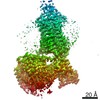
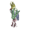
- PDB-7f1t: Crystal structure of the human chemokine receptor CCR5 in complex... -
+ Open data

- Basic information
| Entry | Database: PDB / ID: 7f1t | |||||||||
|---|---|---|---|---|---|---|---|---|---|---|
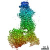
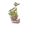
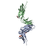
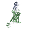
| Title | Crystal structure of the human chemokine receptor CCR5 in complex with MIP-1a | |||||||||
 Components Components | C-C motif chemokine 3,C-C chemokine receptor type 5,Rubredoxin,C-C chemokine receptor type 5 | |||||||||
 Keywords Keywords | SIGNALING PROTEIN / G protein-coupled Receptor / Chemokine receptor CCR5 / MIP-1a | |||||||||
| Function / homology |  Function and homology information Function and homology informationchemokine (C-C motif) ligand 5 binding / lymphocyte chemotaxis / granulocyte chemotaxis / CCR1 chemokine receptor binding / positive regulation of natural killer cell chemotaxis / positive regulation of microglial cell migration / negative regulation of macrophage apoptotic process / astrocyte cell migration / regulation of behavior / alkane catabolic process ...chemokine (C-C motif) ligand 5 binding / lymphocyte chemotaxis / granulocyte chemotaxis / CCR1 chemokine receptor binding / positive regulation of natural killer cell chemotaxis / positive regulation of microglial cell migration / negative regulation of macrophage apoptotic process / astrocyte cell migration / regulation of behavior / alkane catabolic process / CCR5 chemokine receptor binding / eosinophil degranulation / chemokine receptor activity / CCR chemokine receptor binding / regulation of sensory perception of pain / positive regulation of microglial cell activation / signaling / negative regulation of bone mineralization / phosphatidylinositol-4,5-bisphosphate phospholipase C activity / cell activation / T cell chemotaxis / C-C chemokine binding / positive regulation of calcium ion transport / eosinophil chemotaxis / response to cholesterol / C-C chemokine receptor activity / chemokine activity / release of sequestered calcium ion into cytosol by sarcoplasmic reticulum / Chemokine receptors bind chemokines / negative regulation of osteoclast differentiation / dendritic cell chemotaxis / phospholipase activator activity / chemoattractant activity / positive regulation of calcium ion import / Interleukin-10 signaling / exocytosis / macrophage chemotaxis / monocyte chemotaxis / host-mediated suppression of viral transcription / Binding and entry of HIV virion / cellular response to interleukin-1 / cellular defense response / coreceptor activity / neutrophil chemotaxis / positive regulation of calcium-mediated signaling / cytoskeleton organization / positive regulation of interleukin-1 beta production / chemokine-mediated signaling pathway / calcium-mediated signaling / cell chemotaxis / cellular response to type II interferon / cellular response to tumor necrosis factor / response to toxic substance / intracellular calcium ion homeostasis / chemotaxis / osteoblast differentiation / kinase activity / calcium ion transport / positive regulation of inflammatory response / positive regulation of tumor necrosis factor production / MAPK cascade / positive regulation of neuron apoptotic process / cell-cell signaling / regulation of cell shape / antimicrobial humoral immune response mediated by antimicrobial peptide / positive regulation of cytosolic calcium ion concentration / cellular response to lipopolysaccharide / virus receptor activity / actin binding / G alpha (i) signalling events / positive regulation of ERK1 and ERK2 cascade / protein kinase activity / positive regulation of phosphatidylinositol 3-kinase/protein kinase B signal transduction / electron transfer activity / cell surface receptor signaling pathway / endosome / immune response / positive regulation of cell migration / iron ion binding / inflammatory response / G protein-coupled receptor signaling pathway / external side of plasma membrane / negative regulation of gene expression / positive regulation of gene expression / cell surface / : / extracellular region / identical protein binding / plasma membrane / cytosol / cytoplasm Similarity search - Function | |||||||||
| Biological species |  Homo sapiens (human) Homo sapiens (human) Clostridium pasteurianum (bacteria) Clostridium pasteurianum (bacteria) | |||||||||
| Method |  X-RAY DIFFRACTION / SYNCHROTRON / MOLECULAR REPLACEMENT / Resolution: 2.6 Å X-RAY DIFFRACTION / SYNCHROTRON / MOLECULAR REPLACEMENT / Resolution: 2.6 Å | |||||||||
 Authors Authors | Zhang, H. / Chen, K. / Tan, Q. / Han, S. / Zhu, Y. / Zhao, Q. / Wu, B. | |||||||||
| Funding support |  China, 2items China, 2items
| |||||||||
 Citation Citation | Journal: Nat Commun / Year: 2021 Title: Structural basis for chemokine recognition and receptor activation of chemokine receptor CCR5. Authors: Hui Zhang / Kun Chen / Qiuxiang Tan / Qiang Shao / Shuo Han / Chenhui Zhang / Cuiying Yi / Xiaojing Chu / Ya Zhu / Yechun Xu / Qiang Zhao / Beili Wu / Abstract: The chemokine receptor CCR5 plays a vital role in immune surveillance and inflammation. However, molecular details that govern its endogenous chemokine recognition and receptor activation remain ...The chemokine receptor CCR5 plays a vital role in immune surveillance and inflammation. However, molecular details that govern its endogenous chemokine recognition and receptor activation remain elusive. Here we report three cryo-electron microscopy structures of G protein-coupled CCR5 in a ligand-free state and in complex with the chemokine MIP-1α or RANTES, as well as the crystal structure of MIP-1α-bound CCR5. These structures reveal distinct binding modes of the two chemokines and a specific accommodate pattern of the chemokine for the distal N terminus of CCR5. Together with functional data, the structures demonstrate that chemokine-induced rearrangement of toggle switch and plasticity of the receptor extracellular region are critical for receptor activation, while a conserved tryptophan residue in helix II acts as a trigger of receptor constitutive activation. | |||||||||
| History |
|
- Structure visualization
| Structure viewer | Molecule: MolmilJmol/JSmol |
|---|
- Downloads & links
-Download
| PDBx/mmCIF format | 7f1t.cif.gz | 102.6 KB | Display | PDBx/mmCIF format |
|---|---|---|---|---|
| PDB format | pdb7f1t.ent.gz | 75 KB | Display | PDB format |
| PDBx/mmJSON format | 7f1t.json.gz | Tree view | PDBx/mmJSON format | |
| Others |  Other downloads Other downloads |
-Validation report
| Arichive directory | https://data.pdbj.org/pub/pdb/validation_reports/f1/7f1tftp://data.pdbj.org/pub/pdb/validation_reports/f1/7f1t | HTTPS FTP |
|---|
-Related structure data
| Related structure data | 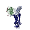 7f1qC  7f1rC 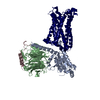 7f1sC 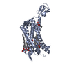 4mbsS S: Starting model for refinement C: citing same article ( |
|---|---|
| Similar structure data |


-Links
 PDBj
PDBj


- Assembly
| Deposited unit | 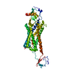
| ||||||||||||
|---|---|---|---|---|---|---|---|---|---|---|---|---|---|
| 1 |
| ||||||||||||
| Unit cell |
|
-Components
| #1: Protein | Mass: 55445.453 Da / Num. of mol.: 1 Mutation: T15C,T108C,C150Y,M156A,G255N,A376D,R417A,T427A,K446E Source method: isolated from a genetically manipulated source Details: Fusion protein of MIP-1a and CCR5 from Homo sapiens, inserted with Rubredoxin from Clostridium pasteurianum Source: (gene. exp.) Homo sapiens (human), (gene. exp.) Clostridium pasteurianum (bacteria)Gene: CCL3, G0S19-1, MIP1A, SCYA3, CCR5, CMKBR5 / Production host:   Spodoptera frugiperda (fall armyworm) Spodoptera frugiperda (fall armyworm)References: UniProt: P10147, UniProt: P51681, UniProt: P00268 |
|---|---|
| #2: Chemical | ChemComp-ZN /   Mass: 65.409 Da / Num. of mol.: 1 / Source method: obtained synthetically / Formula: Zn Mass: 65.409 Da / Num. of mol.: 1 / Source method: obtained synthetically / Formula: Zn |
| Has ligand of interest | N |
| Has protein modification | Y |
-Experimental details
-Experiment
| Experiment | Method: X-RAY DIFFRACTION / Number of used crystals: 1 |
|---|
- Sample preparation
| Crystal | Density Matthews: 3.01 Å3/Da / Density % sol: 64.24 % |
|---|---|
| Crystal grow | Temperature: 293 K / Method: lipidic cubic phase Details: 100mM HEPES, pH 6.0, 250mM ammonium sulfate, 30% (v/v) PEG 400, 8% (v/v) PPG 400 |
-Data collection
| Diffraction | Mean temperature: 100 K / Serial crystal experiment: N |
|---|---|
| Diffraction source | Source: SYNCHROTRON / Site: SPring-8  / Beamline: BL41XU / Wavelength: 1 Å / Beamline: BL41XU / Wavelength: 1 Å |
| Detector | Type: DECTRIS EIGER2 X 16M / Detector: PIXEL / Date: Jun 29, 2018 |
| Radiation | Protocol: SINGLE WAVELENGTH / Monochromatic (M) / Laue (L): M / Scattering type: x-ray |
| Radiation wavelength | Wavelength: 1 Å / Relative weight: 1 |
| Reflection | Resolution: 2.6→50 Å / Num. obs: 18209 / % possible obs: 94.9 % / Redundancy: 6.7 % / CC1/2: 0.985 / Net I/σ(I): 31.7 |
| Reflection shell | Resolution: 2.6→2.64 Å / Num. unique obs: 888 / CC1/2: 0.808 |
- Processing
| Software |
| ||||||||||||||||||||||||
|---|---|---|---|---|---|---|---|---|---|---|---|---|---|---|---|---|---|---|---|---|---|---|---|---|---|
| Refinement | Method to determine structure: MOLECULAR REPLACEMENT Starting model: 4MBS Resolution: 2.6→50 Å / Cross valid method: THROUGHOUT Stereochemistry target values: GeoStd + Monomer Library + CDL v1.2
| ||||||||||||||||||||||||
| Displacement parameters | Biso mean: 117.51 Å2 | ||||||||||||||||||||||||
| Refinement step | Cycle: LAST / Resolution: 2.6→50 Å
| ||||||||||||||||||||||||
| Refine LS restraints |
| ||||||||||||||||||||||||
| LS refinement shell | Resolution: 2.6→2.64 Å
|
