Movie
Movie Controller

[English] 日本語
 Yorodumi
- PDB-7f0l: STRUCTURE OF PHOTOSYNTHETIC LH1-RC SUPER-COMPLEX OF RHODOBACTER S... -
+ Open data

- Basic information
| Entry | Database: PDB / ID: 7f0l | ||||||||||||||||||||||||||||||||||||
|---|---|---|---|---|---|---|---|---|---|---|---|---|---|---|---|---|---|---|---|---|---|---|---|---|---|---|---|---|---|---|---|---|---|---|---|---|---|
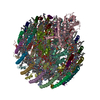
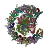
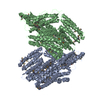
| Title | STRUCTURE OF PHOTOSYNTHETIC LH1-RC SUPER-COMPLEX OF RHODOBACTER SPHAEROIDES MONOMER | ||||||||||||||||||||||||||||||||||||
 Components Components |
| ||||||||||||||||||||||||||||||||||||
 Keywords Keywords | PHOTOSYNTHESIS / LH1-RC COMPLEX / PURPLE BACTERIA | ||||||||||||||||||||||||||||||||||||
| Function / homology |  Function and homology information Function and homology informationorganelle inner membrane / plasma membrane light-harvesting complex / bacteriochlorophyll binding / photosynthesis, light reaction / membrane => GO:0016020 / metal ion binding / plasma membrane Similarity search - Function | ||||||||||||||||||||||||||||||||||||
| Biological species |  Rhodobacter sphaeroides (bacteria) Rhodobacter sphaeroides (bacteria) | ||||||||||||||||||||||||||||||||||||
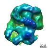
| Method | ELECTRON MICROSCOPY / single particle reconstruction / cryo EM / Resolution: 2.94 Å | ||||||||||||||||||||||||||||||||||||
 Authors Authors | Tani, K. / Nagashima, V.P. / Kanno, R. / Kawamura, S. / Kikuchi, R. / Ji, X.-C. / Hall, M. / Yu, L.-J. / Kimura, Y. / Madigan, M.T. ...Tani, K. / Nagashima, V.P. / Kanno, R. / Kawamura, S. / Kikuchi, R. / Ji, X.-C. / Hall, M. / Yu, L.-J. / Kimura, Y. / Madigan, M.T. / Mizoguchi, A. / Humbel, B.M. / Wang-Otomo, Z.-Y. | ||||||||||||||||||||||||||||||||||||
| Funding support |  Japan, 6items Japan, 6items
| ||||||||||||||||||||||||||||||||||||
 Citation Citation | Journal: Nat Commun / Year: 2021 Title: A previously unrecognized membrane protein in the Rhodobacter sphaeroides LH1-RC photocomplex. Authors: Kazutoshi Tani / Kenji V P Nagashima / Ryo Kanno / Saki Kawamura / Riku Kikuchi / Malgorzata Hall / Long-Jiang Yu / Yukihiro Kimura / Michael T Madigan / Akira Mizoguchi / Bruno M Humbel / ...Authors: Kazutoshi Tani / Kenji V P Nagashima / Ryo Kanno / Saki Kawamura / Riku Kikuchi / Malgorzata Hall / Long-Jiang Yu / Yukihiro Kimura / Michael T Madigan / Akira Mizoguchi / Bruno M Humbel / Zheng-Yu Wang-Otomo /   Abstract: Rhodobacter (Rba.) sphaeroides is the most widely used model organism in bacterial photosynthesis. The light-harvesting-reaction center (LH1-RC) core complex of this purple phototroph is ...Rhodobacter (Rba.) sphaeroides is the most widely used model organism in bacterial photosynthesis. The light-harvesting-reaction center (LH1-RC) core complex of this purple phototroph is characterized by the co-existence of monomeric and dimeric forms, the presence of the protein PufX, and approximately two carotenoids per LH1 αβ-polypeptides. Despite many efforts, structures of the Rba. sphaeroides LH1-RC have not been obtained at high resolutions. Here we report a cryo-EM structure of the monomeric LH1-RC from Rba. sphaeroides strain IL106 at 2.9 Å resolution. The LH1 complex forms a C-shaped structure composed of 14 αβ-polypeptides around the RC with a large ring opening. From the cryo-EM density map, a previously unrecognized integral membrane protein, referred to as protein-U, was identified. Protein-U has a U-shaped conformation near the LH1-ring opening and was annotated as a hypothetical protein in the Rba. sphaeroides genome. Deletion of protein-U resulted in a mutant strain that expressed a much-reduced amount of the dimeric LH1-RC, indicating an important role for protein-U in dimerization of the LH1-RC complex. PufX was located opposite protein-U on the LH1-ring opening, and both its position and conformation differed from that of previous reports of dimeric LH1-RC structures obtained at low-resolution. Twenty-six molecules of the carotenoid spheroidene arranged in two distinct configurations were resolved in the Rba. sphaeroides LH1 and were positioned within the complex to block its channels. Our findings offer an exciting new view of the core photocomplex of Rba. sphaeroides and the connections between structure and function in bacterial photocomplexes in general. | ||||||||||||||||||||||||||||||||||||
| History |
|
- Structure visualization
| Movie |
Movie viewer |
|---|---|
| Structure viewer | Molecule: MolmilJmol/JSmol |
- Downloads & links
-Download
| PDBx/mmCIF format | 7f0l.cif.gz | 518.9 KB | Display | PDBx/mmCIF format |
|---|---|---|---|---|
| PDB format | pdb7f0l.ent.gz | 448.1 KB | Display | PDB format |
| PDBx/mmJSON format | 7f0l.json.gz | Tree view | PDBx/mmJSON format | |
| Others |  Other downloads Other downloads |
-Validation report
| Arichive directory | https://data.pdbj.org/pub/pdb/validation_reports/f0/7f0lftp://data.pdbj.org/pub/pdb/validation_reports/f0/7f0l | HTTPS FTP |
|---|
-Related structure data
| Related structure data |  31400MC M: map data used to model this data C: citing same article ( |
|---|---|
| Similar structure data |


-Links
 PDBj
PDBj

- Assembly
| Deposited unit | 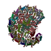
|
|---|---|
| 1 |
|
-Components
-Protein , 3 types, 3 molecules LXU
| #1: Protein | Mass: 31491.611 Da / Num. of mol.: 1 / Source method: isolated from a natural source / Source: (natural) Rhodobacter sphaeroides (bacteria) |
|---|---|
| #7: Protein | Mass: 9017.590 Da / Num. of mol.: 1 / Source method: isolated from a natural source / Source: (natural) Rhodobacter sphaeroides (bacteria) / References: UniProt: A0A330HGC2 |
| #8: Protein | Mass: 5555.558 Da / Num. of mol.: 1 / Source method: isolated from a natural source / Source: (natural) Rhodobacter sphaeroides (bacteria) / References: UniProt: A0A3G6WQU3 |
-Reaction center protein ... , 2 types, 2 molecules MH
| #2: Protein | Mass: 34543.762 Da / Num. of mol.: 1 / Source method: isolated from a natural source / Source: (natural) Rhodobacter sphaeroides (bacteria) |
|---|---|
| #3: Protein | Mass: 28091.350 Da / Num. of mol.: 1 / Source method: isolated from a natural source / Details: WP_069330428.1 / Source: (natural) Rhodobacter sphaeroides (bacteria) |
-Light-harvesting protein B-875 alpha ... , 2 types, 14 molecules ADFIKOQSVY1357
| #4: Protein | Mass: 6473.780 Da / Num. of mol.: 12 / Source method: isolated from a natural source / Details: WP_069330428.1 / Source: (natural) Rhodobacter sphaeroides (bacteria)#6: Protein | Mass: 6445.770 Da / Num. of mol.: 2 / Source method: isolated from a natural source / Source: (natural) Rhodobacter sphaeroides (bacteria) / References: UniProt: P0C0X9 |
|---|
-Protein/peptide / Sugars , 2 types, 38 molecules BEGJNPRTWZ2468
| #16: Sugar | ChemComp-LMT /  Type: D-saccharide / Mass: 510.615 Da / Num. of mol.: 24 / Source method: obtained synthetically / Formula: C24H46O11 / Comment: detergent*YM Type: D-saccharide / Mass: 510.615 Da / Num. of mol.: 24 / Source method: obtained synthetically / Formula: C24H46O11 / Comment: detergent*YM#5: Protein/peptide | Mass: 5592.361 Da / Num. of mol.: 14 / Source method: isolated from a natural source / Source: (natural) Rhodobacter sphaeroides (bacteria) / References: UniProt: Q7B300 |
|---|
-Non-polymers , 8 types, 88 molecules 


| #9: Chemical | ChemComp-BCL /  Mass: 911.504 Da / Num. of mol.: 32 / Source method: obtained synthetically / Formula: C55H74MgN4O6 / Feature type: SUBJECT OF INVESTIGATION Mass: 911.504 Da / Num. of mol.: 32 / Source method: obtained synthetically / Formula: C55H74MgN4O6 / Feature type: SUBJECT OF INVESTIGATION#10: Chemical |  Mass: 889.215 Da / Num. of mol.: 2 / Source method: obtained synthetically / Formula: C55H76N4O6 Mass: 889.215 Da / Num. of mol.: 2 / Source method: obtained synthetically / Formula: C55H76N4O6#11: Chemical |  Mass: 863.343 Da / Num. of mol.: 3 / Source method: obtained synthetically / Formula: C59H90O4 Mass: 863.343 Da / Num. of mol.: 3 / Source method: obtained synthetically / Formula: C59H90O4#12: Chemical | ChemComp-PGV / (  Mass: 749.007 Da / Num. of mol.: 13 / Source method: obtained synthetically / Formula: C40H77O10P / Comment: phospholipid*YM Mass: 749.007 Da / Num. of mol.: 13 / Source method: obtained synthetically / Formula: C40H77O10P / Comment: phospholipid*YM#13: Chemical | ChemComp-LDA /  Mass: 229.402 Da / Num. of mol.: 6 / Source method: obtained synthetically / Formula: C14H31NO / Comment: LDAO, detergent*YM Mass: 229.402 Da / Num. of mol.: 6 / Source method: obtained synthetically / Formula: C14H31NO / Comment: LDAO, detergent*YM#14: Chemical | ChemComp-FE / |  Mass: 55.845 Da / Num. of mol.: 1 / Source method: obtained synthetically / Formula: Fe Mass: 55.845 Da / Num. of mol.: 1 / Source method: obtained synthetically / Formula: Fe#15: Chemical | ChemComp-SPO /  Mass: 568.914 Da / Num. of mol.: 27 / Source method: obtained synthetically / Formula: C41H60O / Feature type: SUBJECT OF INVESTIGATION Mass: 568.914 Da / Num. of mol.: 27 / Source method: obtained synthetically / Formula: C41H60O / Feature type: SUBJECT OF INVESTIGATION#17: Chemical | ChemComp-CDL /  Mass: 1464.043 Da / Num. of mol.: 4 / Source method: obtained synthetically / Formula: C81H156O17P2 / Comment: phospholipid*YM Mass: 1464.043 Da / Num. of mol.: 4 / Source method: obtained synthetically / Formula: C81H156O17P2 / Comment: phospholipid*YM |
|---|
-Details
| Has ligand of interest | Y |
|---|---|
| Has protein modification | Y |
-Experimental details
-Experiment
| Experiment | Method: ELECTRON MICROSCOPY |
|---|---|
| EM experiment | Aggregation state: PARTICLE / 3D reconstruction method: single particle reconstruction |
- Sample preparation
| Component | Name: Photosynthetic LH1-RC complex from the purple phototrophic bacterium Rhodobacter sphaeroides monomer Type: COMPLEX / Entity ID: #1-#8 / Source: NATURAL |
|---|---|
| Molecular weight | Units: MEGADALTONS / Experimental value: NO |
| Source (natural) | Organism: Rhodobacter sphaeroides f. sp. denitrificans (bacteria) Strain: IL106 |
| Buffer solution | pH: 8 |
| Specimen | Conc.: 3 mg/ml / Embedding applied: NO / Shadowing applied: NO / Staining applied: NO / Vitrification applied: YES / Details: This sample was monodisperse. |
| Specimen support | Grid material: MOLYBDENUM |
| Vitrification | Instrument: LEICA EM GP / Cryogen name: ETHANE / Humidity: 80 % / Chamber temperature: 277 K |
- Electron microscopy imaging
| Experimental equipment |  Model: Talos Arctica / Image courtesy: FEI Company |
|---|---|
| Microscopy | Model: FEI TALOS ARCTICA |
| Electron gun | Electron source:  FIELD EMISSION GUN / Accelerating voltage: 200 kV / Illumination mode: FLOOD BEAM FIELD EMISSION GUN / Accelerating voltage: 200 kV / Illumination mode: FLOOD BEAM |
| Electron lens | Mode: BRIGHT FIELD / Alignment procedure: COMA FREE |
| Specimen holder | Cryogen: NITROGEN / Specimen holder model: FEI TITAN KRIOS AUTOGRID HOLDER |
| Image recording | Average exposure time: 1.275 sec. / Electron dose: 42 e/Å2 / Detector mode: COUNTING / Film or detector model: FEI FALCON III (4k x 4k) |
- Processing
| Software | Name: PHENIX / Version: 1.19.2_4158: / Classification: refinement | ||||||||||||||||||||||||||||||||
|---|---|---|---|---|---|---|---|---|---|---|---|---|---|---|---|---|---|---|---|---|---|---|---|---|---|---|---|---|---|---|---|---|---|
| EM software |
| ||||||||||||||||||||||||||||||||
| CTF correction | Type: PHASE FLIPPING ONLY | ||||||||||||||||||||||||||||||||
| Particle selection | Num. of particles selected: 551846 | ||||||||||||||||||||||||||||||||
| 3D reconstruction | Resolution: 2.94 Å / Resolution method: FSC 0.143 CUT-OFF / Num. of particles: 160448 / Symmetry type: POINT | ||||||||||||||||||||||||||||||||
| Atomic model building | B value: 60 / Protocol: RIGID BODY FIT / Space: REAL / Target criteria: Correlation coefficient | ||||||||||||||||||||||||||||||||
| Atomic model building | PDB-ID: 5Y5S Accession code: 5Y5S / Source name: PDB / Type: experimental model | ||||||||||||||||||||||||||||||||
| Refine LS restraints |
|