 Movie
Movie Controller

[English] 日本語
 Yorodumi
- PDB-7ezz: Crystal structure of Salmonella typhi outer membrane phospholipas... -
+ Open data

- Basic information
| Entry | Database: PDB / ID: 7ezz | |||||||||||||||
|---|---|---|---|---|---|---|---|---|---|---|---|---|---|---|---|---|
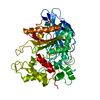
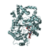
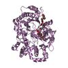
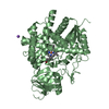
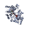
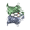
| Title | Crystal structure of Salmonella typhi outer membrane phospholipase (OMPLA) dimer with bound calcium | |||||||||||||||
 Components Components | Phospholipase A1 | |||||||||||||||
 Keywords Keywords | HYDROLASE / Outer membrane phospholipase / OMPLA / Outer membrane protein / Calcium-binding / beta-barrel / MEMBRANE PROTEIN | |||||||||||||||
| Function / homology |  Function and homology information Function and homology information: / : / phospholipase A1 / glycerophospholipid phospholipase A1 activity / A2-type glycerophospholipase activity / phospholipase A2 / lipid catabolic process / cell outer membrane / metal ion binding Similarity search - Function | |||||||||||||||
| Biological species |  Salmonella typhi (bacteria) Salmonella typhi (bacteria) | |||||||||||||||
| Method |  X-RAY DIFFRACTION / MOLECULAR REPLACEMENT / Resolution: 2.76 Å X-RAY DIFFRACTION / MOLECULAR REPLACEMENT / Resolution: 2.76 Å | |||||||||||||||
 Authors Authors | Perumal, P. / Raina, R. / Sreeshma, N.S. / Arockiasamy, A. / Sundarabaalaji, N. | |||||||||||||||
| Funding support |  India, 4items India, 4items
| |||||||||||||||
 Citation Citation | Journal: To Be Published Title: Crystal structure of Salmonella typhi outer membrane phospholipase (OMPLA) dimer with bound calcium Authors: Perumal, P. / Raina, R. / Sreeshma, N.S. / Arockiasamy, A. / Sundarabaalaji, N. | |||||||||||||||
| History |
|
- Structure visualization
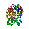
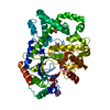
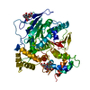
| Structure viewer | Molecule: MolmilJmol/JSmol |
|---|
- Downloads & links
-Download
| PDBx/mmCIF format | 7ezz.cif.gz | 120.3 KB | Display | PDBx/mmCIF format |
|---|---|---|---|---|
| PDB format | pdb7ezz.ent.gz | 90.3 KB | Display | PDB format |
| PDBx/mmJSON format | 7ezz.json.gz | Tree view | PDBx/mmJSON format | |
| Others |  Other downloads Other downloads |
-Validation report
| Arichive directory | https://data.pdbj.org/pub/pdb/validation_reports/ez/7ezzftp://data.pdbj.org/pub/pdb/validation_reports/ez/7ezz | HTTPS FTP |
|---|
-Related structure data
| Related structure data | 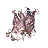 1qd6S S: Starting model for refinement |
|---|---|
| Similar structure data |

-Links
 PDBj
PDBj

- Assembly
| Deposited unit | 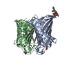
| |||||||||||||||||||||
|---|---|---|---|---|---|---|---|---|---|---|---|---|---|---|---|---|---|---|---|---|---|---|
| 1 |
| |||||||||||||||||||||
| Unit cell |
| |||||||||||||||||||||
| Noncrystallographic symmetry (NCS) | NCS domain:
NCS domain segments: Ens-ID: 1 / Beg auth comp-ID: ALA / Beg label comp-ID: ALA / End auth comp-ID: PHE / End label comp-ID: PHE / Auth seq-ID: 33 - 289 / Label seq-ID: 1 - 257
|
-Components
| #1: Protein | Mass: 29566.723 Da / Num. of mol.: 2 Source method: isolated from a genetically manipulated source Source: (gene. exp.) Salmonella typhi (bacteria) / Gene: pldA, STY3602, t3340 / Production host: References: UniProt: P0A232, phospholipase A1, phospholipase A2 #2: Sugar | ChemComp-BOG /   Type: D-saccharide / Mass: 292.369 Da / Num. of mol.: 4 / Source method: obtained synthetically / Formula: C14H28O6 / Comment: detergent*YM Type: D-saccharide / Mass: 292.369 Da / Num. of mol.: 4 / Source method: obtained synthetically / Formula: C14H28O6 / Comment: detergent*YM#3: Chemical |   Mass: 40.078 Da / Num. of mol.: 2 / Source method: obtained synthetically / Formula: Ca / Feature type: SUBJECT OF INVESTIGATION Mass: 40.078 Da / Num. of mol.: 2 / Source method: obtained synthetically / Formula: Ca / Feature type: SUBJECT OF INVESTIGATION#4: Chemical |   Mass: 170.335 Da / Num. of mol.: 2 / Source method: obtained synthetically / Formula: C12H26 / Feature type: SUBJECT OF INVESTIGATION Mass: 170.335 Da / Num. of mol.: 2 / Source method: obtained synthetically / Formula: C12H26 / Feature type: SUBJECT OF INVESTIGATION#5: Water | ChemComp-HOH / |  Mass: 18.015 Da / Num. of mol.: 54 / Source method: isolated from a natural source / Formula: H2O Mass: 18.015 Da / Num. of mol.: 54 / Source method: isolated from a natural source / Formula: H2OHas ligand of interest | Y | |
|---|
-Experimental details
-Experiment
| Experiment | Method: X-RAY DIFFRACTION / Number of used crystals: 1 |
|---|
- Sample preparation
| Crystal | Density Matthews: 2.57 Å3/Da / Density % sol: 52.18 % |
|---|---|
| Crystal grow | Temperature: 293.15 K / Method: vapor diffusion, sitting drop / pH: 7 Details: 0.1 M sodium iodide, 0.1 M sodium phosphate (pH 7.0), and 33% v/v PEG 300 |
-Data collection
| Diffraction | Mean temperature: 100 K / Serial crystal experiment: N | ||||||||||||||||||||||||||||||
|---|---|---|---|---|---|---|---|---|---|---|---|---|---|---|---|---|---|---|---|---|---|---|---|---|---|---|---|---|---|---|---|
| Diffraction source | Source: ROTATING ANODE / Type: RIGAKU FR-E+ SUPERBRIGHT / Wavelength: 1.54 Å | ||||||||||||||||||||||||||||||
| Detector | Type: RIGAKU RAXIS IV++ / Detector: IMAGE PLATE / Date: Jan 5, 2014 | ||||||||||||||||||||||||||||||
| Radiation | Protocol: SINGLE WAVELENGTH / Monochromatic (M) / Laue (L): M / Scattering type: x-ray | ||||||||||||||||||||||||||||||
| Radiation wavelength | Wavelength: 1.54 Å / Relative weight: 1 | ||||||||||||||||||||||||||||||
| Reflection | Resolution: 2.654→62.879 Å / Num. obs: 17368 / % possible obs: 95.6 % / Redundancy: 6.3 % / CC1/2: 0.991 / Rmerge(I) obs: 0.249 / Rpim(I) all: 0.106 / Rrim(I) all: 0.271 / Net I/σ(I): 7.4 / Num. measured all: 109007 | ||||||||||||||||||||||||||||||
| Reflection shell | Diffraction-ID: 1
|
- Processing
| Software |
| |||||||||||||||||||||||||||||||||||||||||||||
|---|---|---|---|---|---|---|---|---|---|---|---|---|---|---|---|---|---|---|---|---|---|---|---|---|---|---|---|---|---|---|---|---|---|---|---|---|---|---|---|---|---|---|---|---|---|---|
| Refinement | Method to determine structure: MOLECULAR REPLACEMENT Starting model: 1QD6 Resolution: 2.76→47.87 Å / Cor.coef. Fo:Fc: 0.907 / Cor.coef. Fo:Fc free: 0.851 / SU B: 16.311 / SU ML: 0.323 / Cross valid method: THROUGHOUT / σ(F): 0 / ESU R Free: 0.391 / Stereochemistry target values: MAXIMUM LIKELIHOOD / Details: U VALUES : REFINED INDIVIDUALLY
| |||||||||||||||||||||||||||||||||||||||||||||
| Solvent computation | Ion probe radii: 0.8 Å / Shrinkage radii: 0.8 Å / VDW probe radii: 1.2 Å / Solvent model: MASK | |||||||||||||||||||||||||||||||||||||||||||||
| Displacement parameters | Biso max: 91.73 Å2 / Biso mean: 29.007 Å2 / Biso min: 5.53 Å2
| |||||||||||||||||||||||||||||||||||||||||||||
| Refinement step | Cycle: final / Resolution: 2.76→47.87 Å
| |||||||||||||||||||||||||||||||||||||||||||||
| Refine LS restraints |
| |||||||||||||||||||||||||||||||||||||||||||||
| Refine LS restraints NCS | Ens-ID: 1 / Number: 8177 / Refine-ID: X-RAY DIFFRACTION / Type: interatomic distance / Rms dev position: 0.11 Å / Weight position: 0.05
| |||||||||||||||||||||||||||||||||||||||||||||
| LS refinement shell | Resolution: 2.762→2.833 Å / Rfactor Rfree error: 0 / Total num. of bins used: 20
|
