 Movie
Movie Controller


+ Open data

- Basic information
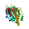
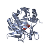
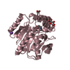
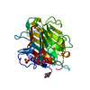
| Entry | Database: PDB / ID: 7ehz | ||||||
|---|---|---|---|---|---|---|---|
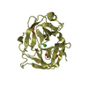
| Title | Structure of human NNMT in complex with macrocyclic peptide 2 | ||||||
 Components Components |
| ||||||
 Keywords Keywords | TRANSFERASE / Nicotinamide N-methyltransferase | ||||||
| Function / homology |  Function and homology information Function and homology informationpyridine N-methyltransferase activity / nicotinamide N-methyltransferase / nicotinamide metabolic process / nicotinamide N-methyltransferase activity / Metabolism of ingested SeMet, Sec, MeSec into H2Se / positive regulation of protein deacetylation / NAD biosynthesis via nicotinamide riboside salvage pathway / Methylation / Nicotinamide salvaging / animal organ regeneration ...pyridine N-methyltransferase activity / nicotinamide N-methyltransferase / nicotinamide metabolic process / nicotinamide N-methyltransferase activity / Metabolism of ingested SeMet, Sec, MeSec into H2Se / positive regulation of protein deacetylation / NAD biosynthesis via nicotinamide riboside salvage pathway / Methylation / Nicotinamide salvaging / animal organ regeneration / positive regulation of gluconeogenesis / : / methylation / response to xenobiotic stimulus / cytosol Similarity search - Function | ||||||
| Biological species |  Homo sapiens (human) Homo sapiens (human)synthetic construct (others) | ||||||
| Method |  X-RAY DIFFRACTION / MOLECULAR REPLACEMENT / molecular replacement / Resolution: 2.5 Å X-RAY DIFFRACTION / MOLECULAR REPLACEMENT / molecular replacement / Resolution: 2.5 Å | ||||||
 Authors Authors | Hayashi, K. / Mikamiyama, H. / Uehara, S. / Yamamoto, S. / Cary, D. / Nishikawa, J. / Ueda, T. / Ozasa, H. / Mihara, K. / Yoshimura, N. ...Hayashi, K. / Mikamiyama, H. / Uehara, S. / Yamamoto, S. / Cary, D. / Nishikawa, J. / Ueda, T. / Ozasa, H. / Mihara, K. / Yoshimura, N. / Kawai, T. / Ono, T. / Yamamoto, S. / Fumoto, M. | ||||||
 Citation Citation | Journal: Acs Med.Chem.Lett. / Year: 2021 Title: Macrocyclic Peptides as a Novel Class of NNMT Inhibitors: A SAR Study Aimed at Inhibitory Activity in the Cell. Authors: Hayashi, K. / Uehara, S. / Yamamoto, S. / Cary, D.R. / Nishikawa, J. / Ueda, T. / Ozasa, H. / Mihara, K. / Yoshimura, N. / Kawai, T. / Ono, T. / Yamamoto, S. / Fumoto, M. / Mikamiyama, H. | ||||||
| History |
|
- Structure visualization
| Structure viewer | Molecule: MolmilJmol/JSmol |
|---|
- Downloads & links
-Download
| PDBx/mmCIF format | 7ehz.cif.gz | 64.8 KB | Display | PDBx/mmCIF format |
|---|---|---|---|---|
| PDB format | pdb7ehz.ent.gz | 43.9 KB | Display | PDB format |
| PDBx/mmJSON format | 7ehz.json.gz | Tree view | PDBx/mmJSON format | |
| Others |  Other downloads Other downloads |
-Validation report
| Summary document | 7ehz_validation.pdf.gz | 442.8 KB | Display | wwPDB validaton report |
|---|---|---|---|---|
| Full document | 7ehz_full_validation.pdf.gz | 447.4 KB | Display | |
| Data in XML | 7ehz_validation.xml.gz | 11.8 KB | Display | |
| Data in CIF | 7ehz_validation.cif.gz | 15.2 KB | Display | |
| Arichive directory | https://data.pdbj.org/pub/pdb/validation_reports/eh/7ehzftp://data.pdbj.org/pub/pdb/validation_reports/eh/7ehz | HTTPS FTP |
-Related structure data
| Related structure data | 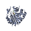 7eguC 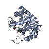 7ei2C 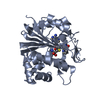 3rodS S: Starting model for refinement C: citing same article ( |
|---|---|
| Similar structure data |


-Links
 PDBj
PDBj- Assembly
| Deposited unit | 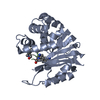
| ||||||||
|---|---|---|---|---|---|---|---|---|---|
| 1 |
| ||||||||
| Unit cell |
|
-Components
| #1: Protein | Mass: 28891.164 Da / Num. of mol.: 1 / Mutation: E103A Source method: isolated from a genetically manipulated source Source: (gene. exp.) Homo sapiens (human) / Gene: NNMT / Production host:  References: UniProt: P40261, nicotinamide N-methyltransferase |
|---|---|
| #2: Protein/peptide | Mass: 1203.391 Da / Num. of mol.: 1 / Source method: obtained synthetically / Source: (synth.) synthetic construct (others) |
| #3: Water | ChemComp-HOH /  Mass: 18.015 Da / Num. of mol.: 24 / Source method: isolated from a natural source / Formula: H2O Mass: 18.015 Da / Num. of mol.: 24 / Source method: isolated from a natural source / Formula: H2O |
| Has ligand of interest | Y |
-Experimental details
-Experiment
| Experiment | Method: X-RAY DIFFRACTION / Number of used crystals: 1 |
|---|
- Sample preparation
| Crystal | Density Matthews: 2.16 Å3/Da / Density % sol: 42.97 % |
|---|---|
| Crystal grow | Temperature: 293 K / Method: vapor diffusion, sitting drop / Details: 0.1M Bicine pH9.0, 20% w/v PEG 6000 |
-Data collection
| Diffraction | Mean temperature: 100 K / Serial crystal experiment: N | |||||||||||||||||||||||||||||||||||||||||||||||||||||||||||||||||||||||||||||||||||||||||||||||||||
|---|---|---|---|---|---|---|---|---|---|---|---|---|---|---|---|---|---|---|---|---|---|---|---|---|---|---|---|---|---|---|---|---|---|---|---|---|---|---|---|---|---|---|---|---|---|---|---|---|---|---|---|---|---|---|---|---|---|---|---|---|---|---|---|---|---|---|---|---|---|---|---|---|---|---|---|---|---|---|---|---|---|---|---|---|---|---|---|---|---|---|---|---|---|---|---|---|---|---|---|---|
| Diffraction source | Source: ROTATING ANODE / Type: RIGAKU FR-E+ SUPERBRIGHT / Wavelength: 1.54178 Å | |||||||||||||||||||||||||||||||||||||||||||||||||||||||||||||||||||||||||||||||||||||||||||||||||||
| Detector | Type: RIGAKU RAXIS VII / Detector: IMAGE PLATE / Date: Mar 7, 2018 | |||||||||||||||||||||||||||||||||||||||||||||||||||||||||||||||||||||||||||||||||||||||||||||||||||
| Radiation | Protocol: SINGLE WAVELENGTH / Monochromatic (M) / Laue (L): M / Scattering type: x-ray | |||||||||||||||||||||||||||||||||||||||||||||||||||||||||||||||||||||||||||||||||||||||||||||||||||
| Radiation wavelength | Wavelength: 1.54178 Å / Relative weight: 1 | |||||||||||||||||||||||||||||||||||||||||||||||||||||||||||||||||||||||||||||||||||||||||||||||||||
| Reflection | Resolution: 2.5→50 Å / Num. obs: 9031 / % possible obs: 99.9 % / Redundancy: 3.6 % / Rmerge(I) obs: 0.066 / Rpim(I) all: 0.04 / Rrim(I) all: 0.077 / Χ2: 1.041 / Net I/σ(I): 13.9 | |||||||||||||||||||||||||||||||||||||||||||||||||||||||||||||||||||||||||||||||||||||||||||||||||||
| Reflection shell | Diffraction-ID: 1
|
-Phasing
| Phasing | Method: molecular replacement | ||||||
|---|---|---|---|---|---|---|---|
| Phasing MR | R rigid body: 0.525
|
- Processing
| Software |
| |||||||||||||||||||||||||||||||||||||||||||||
|---|---|---|---|---|---|---|---|---|---|---|---|---|---|---|---|---|---|---|---|---|---|---|---|---|---|---|---|---|---|---|---|---|---|---|---|---|---|---|---|---|---|---|---|---|---|---|
| Refinement | Method to determine structure: MOLECULAR REPLACEMENT Starting model: 3ROD Resolution: 2.5→50 Å / Cor.coef. Fo:Fc: 0.935 / Cor.coef. Fo:Fc free: 0.928 / SU B: 11.608 / SU ML: 0.259 / SU R Cruickshank DPI: 0.6682 / Cross valid method: THROUGHOUT / σ(F): 0 / ESU R: 0.668 / ESU R Free: 0.304 / Stereochemistry target values: MAXIMUM LIKELIHOOD Details: HYDROGENS HAVE BEEN USED IF PRESENT IN THE INPUT U VALUES : REFINED INDIVIDUALLY
| |||||||||||||||||||||||||||||||||||||||||||||
| Solvent computation | Ion probe radii: 0.8 Å / Shrinkage radii: 0.8 Å / VDW probe radii: 1.2 Å / Solvent model: MASK | |||||||||||||||||||||||||||||||||||||||||||||
| Displacement parameters | Biso max: 115.61 Å2 / Biso mean: 49.802 Å2 / Biso min: 29.11 Å2
| |||||||||||||||||||||||||||||||||||||||||||||
| Refinement step | Cycle: final / Resolution: 2.5→50 Å
| |||||||||||||||||||||||||||||||||||||||||||||
| Refine LS restraints |
| |||||||||||||||||||||||||||||||||||||||||||||
| LS refinement shell | Resolution: 2.5→2.549 Å / Rfactor Rfree error: 0 / Total num. of bins used: 20
|
