 Movie
Movie Controller


+ Open data

- Basic information
| Entry | Database: PDB / ID: 7eep | ||||||
|---|---|---|---|---|---|---|---|
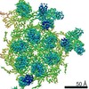
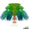
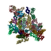
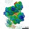
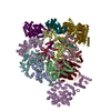
| Title | Cyanophage Pam1 portal-adaptor complex | ||||||
 Components Components |
| ||||||
 Keywords Keywords | VIRUS / Portal and adaptor proteins | ||||||
| Biological species | unidentified (others) | ||||||
| Method | ELECTRON MICROSCOPY / single particle reconstruction / cryo EM / Resolution: 3.75 Å | ||||||
 Authors Authors | Zhang, J.T. / Jiang, Y.L. / Zhou, C.Z. | ||||||
| Funding support |  China, 1items China, 1items
| ||||||
 Citation Citation | Journal: Structure / Year: 2022 Title: Structure and assembly pattern of a freshwater short-tailed cyanophage Pam1. Authors: Jun-Tao Zhang / Feng Yang / Kang Du / Wei-Fang Li / Yuxing Chen / Yong-Liang Jiang / Qiong Li / Cong-Zhao Zhou / Abstract: Despite previous structural analyses of bacteriophages, quite little is known about the structures and assembly patterns of cyanophages. Using cryo-EM combined with crystallography, we solve the near- ...Despite previous structural analyses of bacteriophages, quite little is known about the structures and assembly patterns of cyanophages. Using cryo-EM combined with crystallography, we solve the near-atomic-resolution structure of a freshwater short-tailed cyanophage, Pam1, which comprises a 400-Å-long tail and an icosahedral capsid of 650 Å in diameter. The outer capsid surface is reinforced by trimeric cement proteins with a β-sandwich fold, which structurally resemble the distal motif of Pam1's tailspike, suggesting its potential role in host recognition. At the portal vertex, the dodecameric portal and connected adaptor, followed by a hexameric needle head, form a DNA ejection channel, which is sealed by a trimeric needle. Moreover, we identify a right-handed rifling pattern that might help DNA to revolve along the wall of the ejection channel. Our study reveals the precise assembly pattern of a cyanophage and lays the foundation to support its practical biotechnological and environmental applications. | ||||||
| History |
|
- Structure visualization
| Movie |
 Movie viewer Movie viewer |
|---|---|
| Structure viewer | Molecule: MolmilJmol/JSmol |
- Downloads & links
-Download
| PDBx/mmCIF format | 7eep.cif.gz | 1.4 MB | Display | PDBx/mmCIF format |
|---|---|---|---|---|
| PDB format | pdb7eep.ent.gz | 1.2 MB | Display | PDB format |
| PDBx/mmJSON format | 7eep.json.gz | Tree view | PDBx/mmJSON format | |
| Others |  Other downloads Other downloads |
-Validation report
| Summary document | 7eep_validation.pdf.gz | 1.2 MB | Display | wwPDB validaton report |
|---|---|---|---|---|
| Full document | 7eep_full_validation.pdf.gz | 1.2 MB | Display | |
| Data in XML | 7eep_validation.xml.gz | 200.9 KB | Display | |
| Data in CIF | 7eep_validation.cif.gz | 291.7 KB | Display | |
| Arichive directory | https://data.pdbj.org/pub/pdb/validation_reports/ee/7eepftp://data.pdbj.org/pub/pdb/validation_reports/ee/7eep | HTTPS FTP |
-Related structure data
| Related structure data | 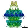 31079MC 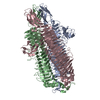 7eeaC 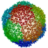 7eelC 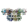 7eeqC M: map data used to model this data C: citing same article ( |
|---|---|
| Similar structure data |

-Links
 PDBj
PDBj- Assembly
| Deposited unit | 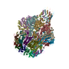
|
|---|---|
| 1 |
|
-Components
| #1: Protein | Mass: 67483.438 Da / Num. of mol.: 12 / Source method: isolated from a natural source / Source: (natural) unidentified (others) #2: Protein | Mass: 19901.504 Da / Num. of mol.: 12 / Source method: isolated from a natural source / Source: (natural) unidentified (others) Source details | The source organism is a short-tailed cyanophage which was separated and sequenced by the author. | |
|---|
-Experimental details
-Experiment
| Experiment | Method: ELECTRON MICROSCOPY |
|---|---|
| EM experiment | Aggregation state: PARTICLE / 3D reconstruction method: single particle reconstruction |
- Sample preparation
| Component | Name: Pam1 / Type: VIRUS / Entity ID: all / Source: NATURAL |
|---|---|
| Source (natural) | Organism: unidentified (others) |
| Details of virus | Empty: NO / Enveloped: NO / Isolate: OTHER / Type: VIRION |
| Natural host | Organism: Pseudanabaena mucicola |
| Buffer solution | pH: 7.5 |
| Specimen | Embedding applied: NO / Shadowing applied: NO / Staining applied: NO / Vitrification applied: YES |
| Vitrification | Cryogen name: ETHANE / Humidity: 100 % / Chamber temperature: 300 K |
- Electron microscopy imaging
| Experimental equipment |  Model: Titan Krios / Image courtesy: FEI Company |
|---|---|
| Microscopy | Model: FEI TITAN KRIOS |
| Electron gun | Electron source:  FIELD EMISSION GUN / Accelerating voltage: 300 kV / Illumination mode: SPOT SCAN FIELD EMISSION GUN / Accelerating voltage: 300 kV / Illumination mode: SPOT SCAN |
| Electron lens | Mode: BRIGHT FIELD |
| Image recording | Electron dose: 50 e/Å2 / Film or detector model: GATAN K2 SUMMIT (4k x 4k) |
- Processing
| EM software | Name: RELION / Version: 3.1 / Category: 3D reconstruction |
|---|---|
| CTF correction | Type: PHASE FLIPPING ONLY |
| 3D reconstruction | Resolution: 3.75 Å / Resolution method: FSC 0.143 CUT-OFF / Num. of particles: 21762 / Symmetry type: POINT |
