 Movie
Movie Controller


+ Open data

- Basic information
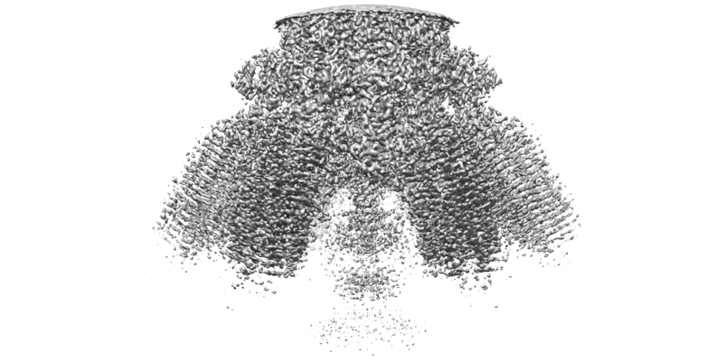
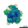
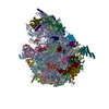
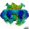
| Entry | Database: EMDB / ID: EMD-31080 | |||||||||
|---|---|---|---|---|---|---|---|---|---|---|
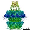
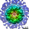
| Title | Cyanophage Pam1 tail machine | |||||||||
 Map data Map data | ||||||||||
 Sample Sample |
| |||||||||
 Keywords Keywords | Needle head proteins and tailspike head-binding proteins / VIRAL PROTEIN | |||||||||
| Biological species | unidentified (others) | |||||||||
| Method | single particle reconstruction / cryo EM / Resolution: 3.96 Å | |||||||||
 Authors Authors | Zhang JT / Jiang YL | |||||||||
| Funding support |  China, 1 items China, 1 items
| |||||||||
 Citation Citation | Journal: Structure / Year: 2022 Title: Structure and assembly pattern of a freshwater short-tailed cyanophage Pam1. Authors: Jun-Tao Zhang / Feng Yang / Kang Du / Wei-Fang Li / Yuxing Chen / Yong-Liang Jiang / Qiong Li / Cong-Zhao Zhou / Abstract: Despite previous structural analyses of bacteriophages, quite little is known about the structures and assembly patterns of cyanophages. Using cryo-EM combined with crystallography, we solve the near- ...Despite previous structural analyses of bacteriophages, quite little is known about the structures and assembly patterns of cyanophages. Using cryo-EM combined with crystallography, we solve the near-atomic-resolution structure of a freshwater short-tailed cyanophage, Pam1, which comprises a 400-Å-long tail and an icosahedral capsid of 650 Å in diameter. The outer capsid surface is reinforced by trimeric cement proteins with a β-sandwich fold, which structurally resemble the distal motif of Pam1's tailspike, suggesting its potential role in host recognition. At the portal vertex, the dodecameric portal and connected adaptor, followed by a hexameric needle head, form a DNA ejection channel, which is sealed by a trimeric needle. Moreover, we identify a right-handed rifling pattern that might help DNA to revolve along the wall of the ejection channel. Our study reveals the precise assembly pattern of a cyanophage and lays the foundation to support its practical biotechnological and environmental applications. | |||||||||
| History |
|
- Structure visualization
| Movie |
 Movie viewer Movie viewer |
|---|---|
| Structure viewer | EM map: SurfViewMolmilJmol/JSmol |
| Supplemental images |
- Downloads & links
-EMDB archive
| Map data | emd_31080.map.gz | 165.7 MB | EMDB map data format | |
|---|---|---|---|---|
| Header (meta data) | emd-31080-v30.xmlemd-31080.xml | 10.3 KB 10.3 KB | Display Display | EMDB header |
| Images |  emd_31080.png emd_31080.png | 161.9 KB | ||
| Filedesc metadata | emd-31080.cif.gz | 5 KB | ||
| Archive directory |  http://ftp.pdbj.org/pub/emdb/structures/EMD-31080ftp://ftp.pdbj.org/pub/emdb/structures/EMD-31080 http://ftp.pdbj.org/pub/emdb/structures/EMD-31080ftp://ftp.pdbj.org/pub/emdb/structures/EMD-31080 | HTTPS FTP |
-Related structure data
| Related structure data | 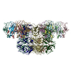 7eeqMC 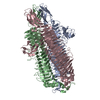 7eeaC 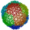 7eelC 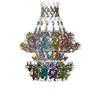 7eepC M: atomic model generated by this map C: citing same article ( |
|---|---|
| Similar structure data |

-Links
| EMDB pages | EMDB (EBI/PDBe) / EMDataResource |
|---|
-Map
| File | Download / File: emd_31080.map.gz / Format: CCP4 / Size: 178 MB / Type: IMAGE STORED AS FLOATING POINT NUMBER (4 BYTES) | ||||||||||||||||||||||||||||||||||||||||||||||||||||||||||||||||||||
|---|---|---|---|---|---|---|---|---|---|---|---|---|---|---|---|---|---|---|---|---|---|---|---|---|---|---|---|---|---|---|---|---|---|---|---|---|---|---|---|---|---|---|---|---|---|---|---|---|---|---|---|---|---|---|---|---|---|---|---|---|---|---|---|---|---|---|---|---|---|
| Projections & slices | Image control
Images are generated by Spider. | ||||||||||||||||||||||||||||||||||||||||||||||||||||||||||||||||||||
| Voxel size | X=Y=Z: 1.013 Å | ||||||||||||||||||||||||||||||||||||||||||||||||||||||||||||||||||||
| Density |
| ||||||||||||||||||||||||||||||||||||||||||||||||||||||||||||||||||||
| Symmetry | Space group: 1 | ||||||||||||||||||||||||||||||||||||||||||||||||||||||||||||||||||||
| Details | EMDB XML:
CCP4 map header:
| ||||||||||||||||||||||||||||||||||||||||||||||||||||||||||||||||||||
 Z (Sec.)
 Y (Row.)
 X (Col.)


-Supplemental data
- Sample components
-Entire : Pam1
| Entire | Name: Pam1 |
|---|---|
| Components |
|
-Supramolecule #1: Pam1
| Supramolecule | Name: Pam1 / type: complex / ID: 1 / Parent: 0 / Macromolecule list: all |
|---|---|
| Source (natural) | Organism: unidentified (others) |
-Macromolecule #1: Needle head proteins
| Macromolecule | Name: Needle head proteins / type: protein_or_peptide / ID: 1 / Number of copies: 6 / Enantiomer: LEVO |
|---|---|
| Source (natural) | Organism: unidentified (others) |
| Molecular weight | Theoretical: 48.534621 KDa |
| Sequence | String: MKIPYGLGAY TRNRGNLPPL ELINLFVEKS DSQGVILQSR KALVEVADVG AGPVRATFQK DGVFGGDRFT LSGDEFYRGA TLLGTVAGG GQARIVSNGL EVLVNAGGLV YSYNGTNFIN AGFPEAAATT IAFTGRYFIG LSAGTGEWYF SAVNNGRSWD A LDFATAEN ...String: MKIPYGLGAY TRNRGNLPPL ELINLFVEKS DSQGVILQSR KALVEVADVG AGPVRATFQK DGVFGGDRFT LSGDEFYRGA TLLGTVAGG GQARIVSNGL EVLVNAGGLV YSYNGTNFIN AGFPEAAATT IAFTGRYFIG LSAGTGEWYF SAVNNGRSWD A LDFATAEN EPDALLDVLV LDGVLVFFGT ESIEFWGFTG DADLPYSPIQ QRVFEQGIYA TGCAVRVDNS FYWVGKDKIV YR NGDVPQA VSDDGIVEKA EGSTNLTLFV LEDERHKFVC LRGDDFTHPH DVTTGQWCEF KSYGRTNFRA TADFGDDETG KIW AWGGYD DEGIIERLFM AGAALEEATQ IDNIRLTCEV GTTPNLVGIY TDPTLEMRFS YDAGNTWEDW EAETLGAQGK YRQR VEWRA LGMFDDPGAL FQFRITDPVS FRLSDVQANA ATGGRQR |
-Macromolecule #2: Tailspike head-binding domain
| Macromolecule | Name: Tailspike head-binding domain / type: protein_or_peptide / ID: 2 / Number of copies: 18 / Enantiomer: LEVO |
|---|---|
| Source (natural) | Organism: unidentified (others) |
| Molecular weight | Theoretical: 10.782913 KDa |
| Sequence | String: MAAELHITPS RATSSNGLNL DGAKWFFYQT GTTTPQSVYT TAALSVAHSN PVVADAAGKF PAIYFDTTLE YRGVLKTADE ATTIYDIDP INSGILSVLG TSS |
-Experimental details
-Structure determination
| Method | cryo EM |
|---|---|
 Processing Processing | single particle reconstruction |
| Aggregation state | particle |
-Sample preparation
| Buffer | pH: 7.5 |
|---|---|
| Vitrification | Cryogen name: ETHANE / Chamber humidity: 100 % / Chamber temperature: 300 K |
- Electron microscopy
| Microscope | FEI TITAN KRIOS |
|---|---|
| Image recording | Film or detector model: GATAN K2 SUMMIT (4k x 4k) / Average electron dose: 50.0 e/Å2 |
| Electron beam | Acceleration voltage: 300 kV / Electron source:  FIELD EMISSION GUN FIELD EMISSION GUN |
| Electron optics | Illumination mode: SPOT SCAN / Imaging mode: BRIGHT FIELD |
| Experimental equipment |  Model: Titan Krios / Image courtesy: FEI Company |
