Movie
Movie Controller


+ Open data

- Basic information
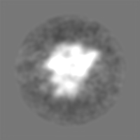
| Entry | Database: EMDB / ID: EMD-5024 | |||||||||
|---|---|---|---|---|---|---|---|---|---|---|
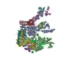
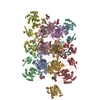
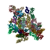
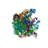
| Title | Cryo-EM structure of Pyrococcus furiosus in apo-state | |||||||||
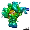
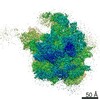
 Map data Map data | Archaea RNAP in its (transcription) elongation state. | |||||||||
 Sample Sample |
| |||||||||
 Keywords Keywords | transcription / initiation / elongation / RNAP / Archaea / cryo-electron microscopy / negative staining / molecular fitting | |||||||||
| Biological species | unidentified (others) | |||||||||
| Method | single particle reconstruction / cryo EM / Resolution: 18.0 Å | |||||||||
 Authors Authors | Carlo SD / Lin S-C / Taatjes D / Hoenger A | |||||||||
 Citation Citation | Journal: Transcription / Year: 2010 Title: Molecular basis of transcription initiation in Archaea. Authors: Sacha De Carlo / Shih-Chieh Lin / Dylan J Taatjes / Andreas Hoenger /  Abstract: Compared with eukaryotes, the archaeal transcription initiation machinery-commonly known as the Pre-Initiation Complex-is relatively simple. The archaeal PIC consists of the TFIIB ortholog TFB, TBP, ...Compared with eukaryotes, the archaeal transcription initiation machinery-commonly known as the Pre-Initiation Complex-is relatively simple. The archaeal PIC consists of the TFIIB ortholog TFB, TBP, and an 11-subunit RNA polymerase (RNAP). The relatively small size of the entire archaeal PIC makes it amenable to structural analysis. Using purified RNAP, TFB, and TBP from the thermophile Pyrococcus furiosus, we assembled the biochemically active PIC at 65ºC. The intact archaeal PIC was isolated by implementing a cross-linking technique followed by size-exclusion chromatography, and the structure of this 440 kDa assembly was determined using electron microscopy and single-particle reconstruction techniques. Combining difference maps with crystal structure docking of various sub-domains, TBP and TFB were localized within the macromolecular PIC. TBP/TFB assemble near the large RpoB subunit and the RpoD/L "foot" domain behind the RNAP central cleft. This location mimics that of yeast TBP and TFIIB in complex with yeast RNAP II. Collectively, these results define the structural organization of the archaeal transcription machinery and suggest a conserved core PIC architecture. | |||||||||
| History |
|
- Structure visualization
| Movie |
 Movie viewer Movie viewer |
|---|---|
| Structure viewer | EM map: SurfViewMolmilJmol/JSmol |
| Supplemental images |

- Downloads & links
-EMDB archive
| Map data | emd_5024.map.gz | 28.3 MB | EMDB map data format | |
|---|---|---|---|---|
| Header (meta data) | emd-5024-v30.xmlemd-5024.xml | 9.2 KB 9.2 KB | Display Display | EMDB header |
| Images | 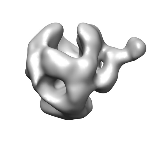 emd_5024_1.jpg emd_5024_1.jpg | 64.2 KB | ||
| Archive directory |  http://ftp.pdbj.org/pub/emdb/structures/EMD-5024ftp://ftp.pdbj.org/pub/emdb/structures/EMD-5024 http://ftp.pdbj.org/pub/emdb/structures/EMD-5024ftp://ftp.pdbj.org/pub/emdb/structures/EMD-5024 | HTTPS FTP |
-Related structure data
-Links
| EMDB pages | EMDB (EBI/PDBe) / EMDataResource |
|---|
-Map
| File | Download / File: emd_5024.map.gz / Format: CCP4 / Size: 29.8 MB / Type: IMAGE STORED AS FLOATING POINT NUMBER (4 BYTES) | ||||||||||||||||||||||||||||||||||||||||||||||||||||||||||||||||||||
|---|---|---|---|---|---|---|---|---|---|---|---|---|---|---|---|---|---|---|---|---|---|---|---|---|---|---|---|---|---|---|---|---|---|---|---|---|---|---|---|---|---|---|---|---|---|---|---|---|---|---|---|---|---|---|---|---|---|---|---|---|---|---|---|---|---|---|---|---|---|
| Annotation | Archaea RNAP in its (transcription) elongation state. | ||||||||||||||||||||||||||||||||||||||||||||||||||||||||||||||||||||
| Projections & slices | Image control
Images are generated by Spider. | ||||||||||||||||||||||||||||||||||||||||||||||||||||||||||||||||||||
| Voxel size | X=Y=Z: 2.25 Å | ||||||||||||||||||||||||||||||||||||||||||||||||||||||||||||||||||||
| Density |
| ||||||||||||||||||||||||||||||||||||||||||||||||||||||||||||||||||||
| Symmetry | Space group: 1 | ||||||||||||||||||||||||||||||||||||||||||||||||||||||||||||||||||||
| Details | EMDB XML:
CCP4 map header:
| ||||||||||||||||||||||||||||||||||||||||||||||||||||||||||||||||||||
 Z (Sec.)
 Y (Row.)
 X (Col.)


-Supplemental data
- Sample components
-Entire : Pyrococcus furiosus RNAP
| Entire | Name: Pyrococcus furiosus RNAP |
|---|---|
| Components |
|
-Supramolecule #1000: Pyrococcus furiosus RNAP
| Supramolecule | Name: Pyrococcus furiosus RNAP / type: sample / ID: 1000 / Oligomeric state: RNAP and DNA / Number unique components: 2 |
|---|---|
| Molecular weight | Experimental: 380 KDa / Method: Mass spectrometry |
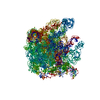
-Macromolecule #1: RNAP apo-state
| Macromolecule | Name: RNAP apo-state / type: protein_or_peptide / ID: 1 / Name.synonym: RNAP elongation complex / Number of copies: 1 / Oligomeric state: multi-subunit complex / Recombinant expression: No / Database: NCBI |
|---|---|
| Source (natural) | Organism: unidentified (others) / Cell: Pyrococcus |
| Molecular weight | Experimental: 380 KDa |
-Experimental details
-Structure determination
| Method | cryo EM |
|---|---|
 Processing Processing | single particle reconstruction |
| Aggregation state | particle |
-Sample preparation
| Concentration | 0.5 mg/mL |
|---|---|
| Buffer | Details: Hepes 0.1 M, pH 8.0, NaCl 50 mM, EDTA 0.1mM, DTT 2 mM |
| Grid | Details: C-Flat |
| Vitrification | Cryogen name: ETHANE / Chamber humidity: 65 % / Instrument: OTHER |
- Electron microscopy
| Microscope | FEI TECNAI F20 |
|---|---|
| Image recording | Category: FILM / Film or detector model: KODAK SO-163 FILM / Digitization - Scanner: NIKON SUPER COOLSCAN 9000 / Digitization - Sampling interval: 6.7 µm / Number real images: 32 / Average electron dose: 15 e/Å2 |
| Electron beam | Acceleration voltage: 200 kV / Electron source:  FIELD EMISSION GUN FIELD EMISSION GUN |
| Electron optics | Calibrated magnification: 49785 / Illumination mode: FLOOD BEAM / Imaging mode: BRIGHT FIELD / Cs: 2.0 mm / Nominal magnification: 50000 |
| Sample stage | Specimen holder: 626 / Specimen holder model: GATAN LIQUID NITROGEN |
| Experimental equipment |  Model: Tecnai F20 / Image courtesy: FEI Company |
-Image processing
| CTF correction | Details: phase-flipped |
|---|---|
| Final reconstruction | Algorithm: OTHER / Resolution.type: BY AUTHOR / Resolution: 18.0 Å / Resolution method: OTHER / Software - Name: SPIDER, EMAN / Number images used: 4611 |
-Atomic model buiding 1
| Initial model | PDB ID: |
|---|---|
| Software | Name: NORMA |
| Details | Protocol: Rigid Body. Manual fitting with CHIMERA first. |
| Refinement | Space: RECIPROCAL / Protocol: RIGID BODY FIT |