 Movie
Movie Controller

[English] 日本語
 Yorodumi
- PDB-7e9g: Cryo-EM structure of Gi-bound metabotropic glutamate receptor mGlu2 -
+ Open data

- Basic information
| Entry | Database: PDB / ID: 7e9g | ||||||||||||||||||
|---|---|---|---|---|---|---|---|---|---|---|---|---|---|---|---|---|---|---|---|
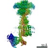
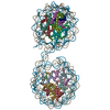
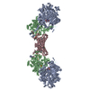
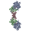
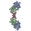
| Title | Cryo-EM structure of Gi-bound metabotropic glutamate receptor mGlu2 | ||||||||||||||||||
 Components Components |
| ||||||||||||||||||
 Keywords Keywords | MEMBRANE PROTEIN / mGlu2 / GPCR / Cryo-EM / complex | ||||||||||||||||||
| Function / homology |  Function and homology information Function and homology informationregulation of response to drug / regulation of glutamate secretion / group II metabotropic glutamate receptor activity / intracellular glutamate homeostasis / behavioral response to nicotine / negative regulation of adenylate cyclase activity / G protein-coupled glutamate receptor signaling pathway / glutamate secretion / Class C/3 (Metabotropic glutamate/pheromone receptors) / glutamate receptor activity ...regulation of response to drug / regulation of glutamate secretion / group II metabotropic glutamate receptor activity / intracellular glutamate homeostasis / behavioral response to nicotine / negative regulation of adenylate cyclase activity / G protein-coupled glutamate receptor signaling pathway / glutamate secretion / Class C/3 (Metabotropic glutamate/pheromone receptors) / glutamate receptor activity / long-term synaptic depression / astrocyte projection / cellular response to stress / regulation of dopamine secretion / regulation of synaptic transmission, glutamatergic / regulation of eating behavior / adenylate cyclase inhibitor activity / positive regulation of protein localization to cell cortex / T cell migration / positive regulation of relaxation of smooth muscle / Adenylate cyclase inhibitory pathway / D2 dopamine receptor binding / adenylate cyclase-inhibiting serotonin receptor signaling pathway / G protein-coupled serotonin receptor binding / presynaptic modulation of chemical synaptic transmission / cellular response to forskolin / mast cell degranulation / regulation of mitotic spindle organization / chemokine-mediated signaling pathway / response to cocaine / calcium channel regulator activity / Regulation of insulin secretion / neuropeptide signaling pathway / response to prostaglandin E / positive regulation of cholesterol biosynthetic process / G protein-coupled receptor binding / response to peptide hormone / G protein-coupled receptor activity / G-protein beta/gamma-subunit complex binding / adenylate cyclase-modulating G protein-coupled receptor signaling pathway / adenylate cyclase-inhibiting G protein-coupled receptor signaling pathway / Olfactory Signaling Pathway / Activation of the phototransduction cascade / G protein-coupled acetylcholine receptor signaling pathway / G beta:gamma signalling through PLC beta / Presynaptic function of Kainate receptors / Thromboxane signalling through TP receptor / Activation of G protein gated Potassium channels / Inhibition of voltage gated Ca2+ channels via Gbeta/gamma subunits / G-protein activation / Glucagon signaling in metabolic regulation / G beta:gamma signalling through CDC42 / Prostacyclin signalling through prostacyclin receptor / Synthesis, secretion, and inactivation of Glucagon-like Peptide-1 (GLP-1) / G beta:gamma signalling through BTK / photoreceptor disc membrane / GDP binding / ADP signalling through P2Y purinoceptor 12 / Glucagon-type ligand receptors / Sensory perception of sweet, bitter, and umami (glutamate) taste / Adrenaline,noradrenaline inhibits insulin secretion / Vasopressin regulates renal water homeostasis via Aquaporins / Glucagon-like Peptide-1 (GLP1) regulates insulin secretion / G alpha (z) signalling events / cellular response to catecholamine stimulus / ADP signalling through P2Y purinoceptor 1 / G beta:gamma signalling through PI3Kgamma / ADORA2B mediated anti-inflammatory cytokines production / adenylate cyclase-activating dopamine receptor signaling pathway / Cooperation of PDCL (PhLP1) and TRiC/CCT in G-protein beta folding / GPER1 signaling / cellular response to prostaglandin E stimulus / heterotrimeric G-protein complex / Inactivation, recovery and regulation of the phototransduction cascade / G alpha (12/13) signalling events / G-protein beta-subunit binding / extracellular vesicle / sensory perception of taste / Thrombin signalling through proteinase activated receptors (PARs) / signaling receptor complex adaptor activity / adenylate cyclase-activating G protein-coupled receptor signaling pathway / retina development in camera-type eye / fibroblast proliferation / gene expression / GTPase binding / presynaptic membrane / scaffold protein binding / G protein activity / midbody / Ca2+ pathway / cell cortex / chemical synaptic transmission / High laminar flow shear stress activates signaling by PIEZO1 and PECAM1:CDH5:KDR in endothelial cells / G alpha (i) signalling events / G alpha (s) signalling events / G alpha (q) signalling events / phospholipase C-activating G protein-coupled receptor signaling pathway / Hydrolases; Acting on acid anhydrides; Acting on GTP to facilitate cellular and subcellular movement / Ras protein signal transduction / positive regulation of phosphatidylinositol 3-kinase/protein kinase B signal transduction Similarity search - Function | ||||||||||||||||||
| Biological species |  Homo sapiens (human) Homo sapiens (human) | ||||||||||||||||||
| Method | ELECTRON MICROSCOPY / single particle reconstruction / cryo EM / Resolution: 3.5 Å | ||||||||||||||||||
 Authors Authors | Lin, S. / Han, S. / Zhao, Q. / Wu, B. | ||||||||||||||||||
| Funding support |  China, 5items China, 5items
| ||||||||||||||||||
 Citation Citation | Journal: Nature / Year: 2021 Title: Structures of G-bound metabotropic glutamate receptors mGlu2 and mGlu4. Authors: Shuling Lin / Shuo Han / Xiaoqing Cai / Qiuxiang Tan / Kexiu Zhou / Dejian Wang / Xinwei Wang / Juan Du / Cuiying Yi / Xiaojing Chu / Antao Dai / Yan Zhou / Yan Chen / Yu Zhou / Hong Liu / ...Authors: Shuling Lin / Shuo Han / Xiaoqing Cai / Qiuxiang Tan / Kexiu Zhou / Dejian Wang / Xinwei Wang / Juan Du / Cuiying Yi / Xiaojing Chu / Antao Dai / Yan Zhou / Yan Chen / Yu Zhou / Hong Liu / Jianfeng Liu / Dehua Yang / Ming-Wei Wang / Qiang Zhao / Beili Wu / Abstract: The metabotropic glutamate receptors (mGlus) have key roles in modulating cell excitability and synaptic transmission in response to glutamate (the main excitatory neurotransmitter in the central ...The metabotropic glutamate receptors (mGlus) have key roles in modulating cell excitability and synaptic transmission in response to glutamate (the main excitatory neurotransmitter in the central nervous system). It has previously been suggested that only one receptor subunit within an mGlu homodimer is responsible for coupling to G protein during receptor activation. However, the molecular mechanism that underlies the asymmetric signalling of mGlus remains unknown. Here we report two cryo-electron microscopy structures of human mGlu2 and mGlu4 bound to heterotrimeric G protein. The structures reveal a G-protein-binding site formed by three intracellular loops and helices III and IV that is distinct from the corresponding binding site in all of the other G-protein-coupled receptor (GPCR) structures. Furthermore, we observed an asymmetric dimer interface of the transmembrane domain of the receptor in the two mGlu-G structures. We confirmed that the asymmetric dimerization is crucial for receptor activation, which was supported by functional data; this dimerization may provide a molecular basis for the asymmetric signal transduction of mGlus. These findings offer insights into receptor signalling of class C GPCRs. | ||||||||||||||||||
| History |
|
- Structure visualization
| Movie |
Movie viewer |
|---|---|
| Structure viewer | Molecule: MolmilJmol/JSmol |
- Downloads & links
-Download
| PDBx/mmCIF format | 7e9g.cif.gz | 477.2 KB | Display | PDBx/mmCIF format |
|---|---|---|---|---|
| PDB format | pdb7e9g.ent.gz | 366.8 KB | Display | PDB format |
| PDBx/mmJSON format | 7e9g.json.gz | Tree view | PDBx/mmJSON format | |
| Others |  Other downloads Other downloads |
-Validation report
| Arichive directory | https://data.pdbj.org/pub/pdb/validation_reports/e9/7e9gftp://data.pdbj.org/pub/pdb/validation_reports/e9/7e9g | HTTPS FTP |
|---|
-Related structure data
| Related structure data | 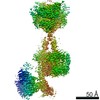 31031MC 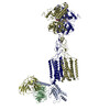 7e9hC M: map data used to model this data C: citing same article ( |
|---|---|
| Similar structure data |


-Links
 PDBj
PDBj


- Assembly
| Deposited unit | 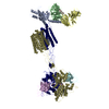
|
|---|---|
| 1 |
|
-Components
-Protein , 1 types, 2 molecules RS
| #1: Protein | Mass: 90240.281 Da / Num. of mol.: 2 / Mutation: S601A Source method: isolated from a genetically manipulated source Source: (gene. exp.) Homo sapiens (human) / Gene: GRM2, GPRC1B, MGLUR2 / Production host:  Baculovirus expression vector pFastBac1-HM / References: UniProt: Q14416 Baculovirus expression vector pFastBac1-HM / References: UniProt: Q14416 |
|---|
-Guanine nucleotide-binding protein ... , 3 types, 3 molecules ABC
| #2: Protein | Mass: 40414.047 Da / Num. of mol.: 1 / Mutation: S47N, G203A, E245A, A326S Source method: isolated from a genetically manipulated source Source: (gene. exp.) Homo sapiens (human) / Gene: GNAI1 / Production host: Baculovirus expression vector pFastBac1-HM / References: UniProt: P63096 |
|---|---|
| #3: Protein | Mass: 38744.371 Da / Num. of mol.: 1 Source method: isolated from a genetically manipulated source Source: (gene. exp.) Homo sapiens (human) / Gene: GNB1 / Production host: Baculovirus expression vector pFastBac1-HM / References: UniProt: P62873 |
| #4: Protein | Mass: 7861.143 Da / Num. of mol.: 1 Source method: isolated from a genetically manipulated source Source: (gene. exp.) Homo sapiens (human) / Gene: GNG2 / Production host: Baculovirus expression vector pFastBac1-HM / References: UniProt: P59768 |
-Antibody , 2 types, 3 molecules DEF
| #5: Antibody | Mass: 27409.588 Da / Num. of mol.: 1 Source method: isolated from a genetically manipulated source Source: (gene. exp.) Homo sapiens (human) / Production host: Baculovirus expression vector pFastBac1-HM |
|---|---|
| #6: Antibody | Mass: 13637.166 Da / Num. of mol.: 2 Source method: isolated from a genetically manipulated source Source: (gene. exp.)  |
-Non-polymers , 2 types, 3 molecules 

| #7: Chemical |  Mass: 185.177 Da / Num. of mol.: 2 / Source method: obtained synthetically / Formula: C8H11NO4 / Feature type: SUBJECT OF INVESTIGATION Mass: 185.177 Da / Num. of mol.: 2 / Source method: obtained synthetically / Formula: C8H11NO4 / Feature type: SUBJECT OF INVESTIGATION#8: Chemical | ChemComp-HZR / |  Mass: 344.878 Da / Num. of mol.: 1 / Source method: obtained synthetically / Formula: C20H25ClN2O / Feature type: SUBJECT OF INVESTIGATION Mass: 344.878 Da / Num. of mol.: 1 / Source method: obtained synthetically / Formula: C20H25ClN2O / Feature type: SUBJECT OF INVESTIGATION |
|---|
-Details
| Has ligand of interest | Y |
|---|---|
| Has protein modification | Y |
-Experimental details
-Experiment
| Experiment | Method: ELECTRON MICROSCOPY |
|---|---|
| EM experiment | Aggregation state: PARTICLE / 3D reconstruction method: single particle reconstruction |
- Sample preparation
| Component | Name: Complex of mGlu2 homodimer with heterotrimeric Gi1 protein in presence of antibodies Type: COMPLEX / Entity ID: #1-#6 / Source: RECOMBINANT |
|---|---|
| Source (natural) | Organism: Homo sapiens (human) |
| Source (recombinant) | Organism: Baculovirus expression vector pFastBac1-HM |
| Buffer solution | pH: 7.4 |
| Specimen | Embedding applied: NO / Shadowing applied: NO / Staining applied: NO / Vitrification applied: YES |
| Vitrification | Cryogen name: ETHANE |
- Electron microscopy imaging
| Experimental equipment |  Model: Titan Krios / Image courtesy: FEI Company |
|---|---|
| Microscopy | Model: FEI TITAN KRIOS |
| Electron gun | Electron source:  FIELD EMISSION GUN / Accelerating voltage: 300 kV / Illumination mode: SPOT SCAN FIELD EMISSION GUN / Accelerating voltage: 300 kV / Illumination mode: SPOT SCAN |
| Electron lens | Mode: BRIGHT FIELD |
| Image recording | Electron dose: 70 e/Å2 / Film or detector model: GATAN K3 BIOQUANTUM (6k x 4k) |
- Processing
| Software |
| ||||||||||||||||||||||||
|---|---|---|---|---|---|---|---|---|---|---|---|---|---|---|---|---|---|---|---|---|---|---|---|---|---|
| CTF correction | Type: PHASE FLIPPING AND AMPLITUDE CORRECTION | ||||||||||||||||||||||||
| 3D reconstruction | Resolution: 3.5 Å / Resolution method: FSC 0.143 CUT-OFF / Num. of particles: 850700 / Symmetry type: POINT | ||||||||||||||||||||||||
| Refinement | Cross valid method: NONE Stereochemistry target values: GeoStd + Monomer Library + CDL v1.2 | ||||||||||||||||||||||||
| Displacement parameters | Biso mean: 37.64 Å2 | ||||||||||||||||||||||||
| Refine LS restraints |
|
