 ムービー
ムービー コントローラー


+ データを開く

- 基本情報
基本情報
| 登録情報 | データベース: EMDB / ID: EMD-31032 | ||||||||||||||||||
|---|---|---|---|---|---|---|---|---|---|---|---|---|---|---|---|---|---|---|---|
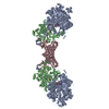
| タイトル | Cryo-EM structure of Gi-bound metabotropic glutamate receptor mGlu4 | ||||||||||||||||||
 マップデータ マップデータ | |||||||||||||||||||
 試料 試料 |
| ||||||||||||||||||
 キーワード キーワード | mGlu4 / GPCR / Cryo-EM / complex / MEMBRANE PROTEIN | ||||||||||||||||||
| 機能・相同性 |  機能・相同性情報 機能・相同性情報adenylate cyclase-inhibiting G protein-coupled glutamate receptor signaling pathway / adenylate cyclase inhibiting G protein-coupled glutamate receptor activity / negative regulation of adenylate cyclase activity / G protein-coupled glutamate receptor signaling pathway / neurotransmitter secretion / Class C/3 (Metabotropic glutamate/pheromone receptors) / GTP metabolic process / glutamate receptor activity / positive regulation of macroautophagy / regulation of synaptic transmission, glutamatergic ...adenylate cyclase-inhibiting G protein-coupled glutamate receptor signaling pathway / adenylate cyclase inhibiting G protein-coupled glutamate receptor activity / negative regulation of adenylate cyclase activity / G protein-coupled glutamate receptor signaling pathway / neurotransmitter secretion / Class C/3 (Metabotropic glutamate/pheromone receptors) / GTP metabolic process / glutamate receptor activity / positive regulation of macroautophagy / regulation of synaptic transmission, glutamatergic / centriolar satellite / Adenylate cyclase inhibitory pathway / regulation of neuron apoptotic process / sperm principal piece / G protein-coupled receptor binding / G protein-coupled receptor activity / G-protein beta/gamma-subunit complex binding / adenylate cyclase-modulating G protein-coupled receptor signaling pathway / adenylate cyclase-inhibiting G protein-coupled receptor signaling pathway / Olfactory Signaling Pathway / Activation of the phototransduction cascade / G protein-coupled acetylcholine receptor signaling pathway / G beta:gamma signalling through PLC beta / Presynaptic function of Kainate receptors / Thromboxane signalling through TP receptor / Activation of G protein gated Potassium channels / Inhibition of voltage gated Ca2+ channels via Gbeta/gamma subunits / G-protein activation / Glucagon signaling in metabolic regulation / G beta:gamma signalling through CDC42 / Prostacyclin signalling through prostacyclin receptor / Synthesis, secretion, and inactivation of Glucagon-like Peptide-1 (GLP-1) / G beta:gamma signalling through BTK / photoreceptor disc membrane / GDP binding / ADP signalling through P2Y purinoceptor 12 / Glucagon-type ligand receptors / Sensory perception of sweet, bitter, and umami (glutamate) taste / Adrenaline,noradrenaline inhibits insulin secretion / Vasopressin regulates renal water homeostasis via Aquaporins / Glucagon-like Peptide-1 (GLP1) regulates insulin secretion / G alpha (z) signalling events / cellular response to catecholamine stimulus / ADP signalling through P2Y purinoceptor 1 / G beta:gamma signalling through PI3Kgamma / ADORA2B mediated anti-inflammatory cytokines production / adenylate cyclase-activating dopamine receptor signaling pathway / Cooperation of PDCL (PhLP1) and TRiC/CCT in G-protein beta folding / GPER1 signaling / cellular response to prostaglandin E stimulus / heterotrimeric G-protein complex / Inactivation, recovery and regulation of the phototransduction cascade / G alpha (12/13) signalling events / G-protein beta-subunit binding / extracellular vesicle / sensory perception of taste / presynapse / Thrombin signalling through proteinase activated receptors (PARs) / signaling receptor complex adaptor activity / retina development in camera-type eye / fibroblast proliferation / GTPase binding / cytoplasmic vesicle / midbody / Ca2+ pathway / chemical synaptic transmission / High laminar flow shear stress activates signaling by PIEZO1 and PECAM1:CDH5:KDR in endothelial cells / G alpha (i) signalling events / G alpha (s) signalling events / G alpha (q) signalling events / phospholipase C-activating G protein-coupled receptor signaling pathway / positive regulation of MAPK cascade / Ras protein signal transduction / cell population proliferation / Extra-nuclear estrogen signaling / ciliary basal body / G protein-coupled receptor signaling pathway / cell division / lysosomal membrane / GTPase activity / centrosome / synapse / nucleolus / GTP binding / protein-containing complex binding / Golgi apparatus / signal transduction / extracellular exosome / nucleoplasm / membrane / metal ion binding / plasma membrane / cytosol / cytoplasm 類似検索 - 分子機能 | ||||||||||||||||||
| 生物種 |  Homo sapiens (ヒト) Homo sapiens (ヒト) | ||||||||||||||||||
| 手法 | 単粒子再構成法 / クライオ電子顕微鏡法 / 解像度: 4.0 Å | ||||||||||||||||||
 データ登録者 データ登録者 | Lin S / Han S | ||||||||||||||||||
| 資金援助 |  中国, 5件 中国, 5件
| ||||||||||||||||||
 引用 引用 | ジャーナル: Nature / 年: 2021 タイトル: Structures of G-bound metabotropic glutamate receptors mGlu2 and mGlu4. 著者: Shuling Lin / Shuo Han / Xiaoqing Cai / Qiuxiang Tan / Kexiu Zhou / Dejian Wang / Xinwei Wang / Juan Du / Cuiying Yi / Xiaojing Chu / Antao Dai / Yan Zhou / Yan Chen / Yu Zhou / Hong Liu / ...著者: Shuling Lin / Shuo Han / Xiaoqing Cai / Qiuxiang Tan / Kexiu Zhou / Dejian Wang / Xinwei Wang / Juan Du / Cuiying Yi / Xiaojing Chu / Antao Dai / Yan Zhou / Yan Chen / Yu Zhou / Hong Liu / Jianfeng Liu / Dehua Yang / Ming-Wei Wang / Qiang Zhao / Beili Wu / 要旨: The metabotropic glutamate receptors (mGlus) have key roles in modulating cell excitability and synaptic transmission in response to glutamate (the main excitatory neurotransmitter in the central ...The metabotropic glutamate receptors (mGlus) have key roles in modulating cell excitability and synaptic transmission in response to glutamate (the main excitatory neurotransmitter in the central nervous system). It has previously been suggested that only one receptor subunit within an mGlu homodimer is responsible for coupling to G protein during receptor activation. However, the molecular mechanism that underlies the asymmetric signalling of mGlus remains unknown. Here we report two cryo-electron microscopy structures of human mGlu2 and mGlu4 bound to heterotrimeric G protein. The structures reveal a G-protein-binding site formed by three intracellular loops and helices III and IV that is distinct from the corresponding binding site in all of the other G-protein-coupled receptor (GPCR) structures. Furthermore, we observed an asymmetric dimer interface of the transmembrane domain of the receptor in the two mGlu-G structures. We confirmed that the asymmetric dimerization is crucial for receptor activation, which was supported by functional data; this dimerization may provide a molecular basis for the asymmetric signal transduction of mGlus. These findings offer insights into receptor signalling of class C GPCRs. | ||||||||||||||||||
| 履歴 |
|
- 構造の表示
構造の表示
| ムービー |
ムービービューア |
|---|---|
| 構造ビューア | EMマップ: SurfViewMolmilJmol/JSmol |
| 添付画像 |

- ダウンロードとリンク
-EMDBアーカイブ
| マップデータ | emd_31032.map.gz | 158.2 MB | EMDBマップデータ形式 | |
|---|---|---|---|---|
| ヘッダ (付随情報) | emd-31032-v30.xmlemd-31032.xml | 16.7 KB 16.7 KB | 表示 表示 | EMDBヘッダ |
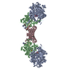
| 画像 | 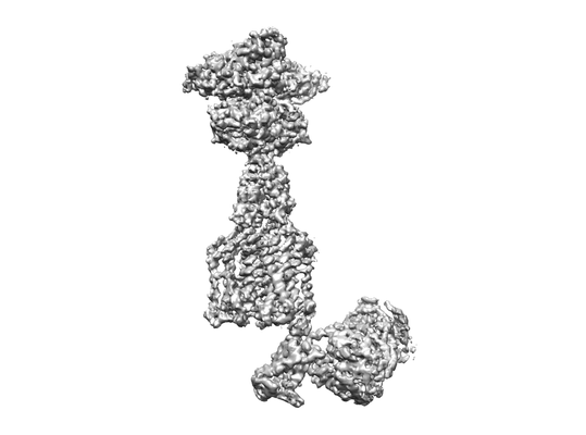 emd_31032.png emd_31032.png | 33.8 KB | ||
| Filedesc metadata | emd-31032.cif.gz | 6.6 KB | ||
| アーカイブディレクトリ |  http://ftp.pdbj.org/pub/emdb/structures/EMD-31032ftp://ftp.pdbj.org/pub/emdb/structures/EMD-31032 http://ftp.pdbj.org/pub/emdb/structures/EMD-31032ftp://ftp.pdbj.org/pub/emdb/structures/EMD-31032 | HTTPS FTP |
-関連構造データ


-リンク
| EMDBのページ | EMDB (EBI/PDBe) / EMDataResource |
|---|---|
| 「今月の分子」の関連する項目 |


-マップ
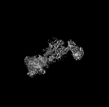
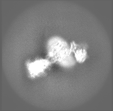
| ファイル | ダウンロード / ファイル: emd_31032.map.gz / 形式: CCP4 / 大きさ: 189.1 MB / タイプ: IMAGE STORED AS FLOATING POINT NUMBER (4 BYTES) | ||||||||||||||||||||||||||||||||||||||||||||||||||||||||||||||||||||
|---|---|---|---|---|---|---|---|---|---|---|---|---|---|---|---|---|---|---|---|---|---|---|---|---|---|---|---|---|---|---|---|---|---|---|---|---|---|---|---|---|---|---|---|---|---|---|---|---|---|---|---|---|---|---|---|---|---|---|---|---|---|---|---|---|---|---|---|---|---|
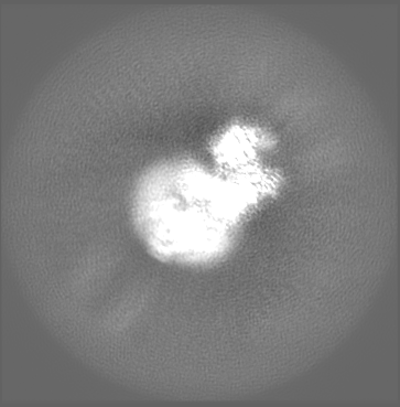
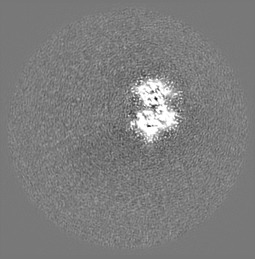
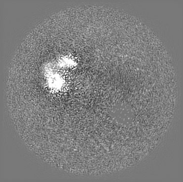
| 投影像・断面図 | 画像のコントロール
画像は Spider により作成 これらの図は立方格子座標系で作成されたものです | ||||||||||||||||||||||||||||||||||||||||||||||||||||||||||||||||||||
| ボクセルのサイズ | X=Y=Z: 1.045 Å | ||||||||||||||||||||||||||||||||||||||||||||||||||||||||||||||||||||
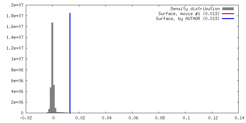
| 密度 |
| ||||||||||||||||||||||||||||||||||||||||||||||||||||||||||||||||||||
| 対称性 | 空間群: 1 | ||||||||||||||||||||||||||||||||||||||||||||||||||||||||||||||||||||
| 詳細 | EMDB XML:
CCP4マップ ヘッダ情報:
| ||||||||||||||||||||||||||||||||||||||||||||||||||||||||||||||||||||
 Z (Sec.)
 Y (Row.)
 X (Col.)


-添付データ
- 試料の構成要素
-全体 : Complex of mGlu4 homodimer with heterotrimeric Gi3 protein in pre...
| 全体 | 名称: Complex of mGlu4 homodimer with heterotrimeric Gi3 protein in presence of antibody |
|---|---|
| 要素 |
|
-超分子 #1: Complex of mGlu4 homodimer with heterotrimeric Gi3 protein in pre...
| 超分子 | 名称: Complex of mGlu4 homodimer with heterotrimeric Gi3 protein in presence of antibody タイプ: complex / ID: 1 / 親要素: 0 / 含まれる分子: #1-#5 |
|---|---|
| 由来(天然) | 生物種: Homo sapiens (ヒト) |
-分子 #1: Metabotropic glutamate receptor 4
| 分子 | 名称: Metabotropic glutamate receptor 4 / タイプ: protein_or_peptide / ID: 1 / コピー数: 2 / 光学異性体: LEVO |
|---|---|
| 由来(天然) | 生物種: Homo sapiens (ヒト) |
| 分子量 | 理論値: 99.458359 KDa |
| 組換発現 | 生物種:  Baculovirus expression vector pFastBac1-HM (ウイルス) Baculovirus expression vector pFastBac1-HM (ウイルス) |
| 配列 | 文字列: DYKDDDDGAP KPKGHPHMNS IRIDGDITLG GLFPVHGRGS EGKPCGELKK EKGIHRLEAM LFALDRINND PDLLPNITLG ARILDTCSR DTHALEQSLT FVQALIEKDG TEVRCGSGGP PIITKPERVV GVIGASGSSV SIMVANILRL FKIPQISYAS T APDLSDNS ...文字列: DYKDDDDGAP KPKGHPHMNS IRIDGDITLG GLFPVHGRGS EGKPCGELKK EKGIHRLEAM LFALDRINND PDLLPNITLG ARILDTCSR DTHALEQSLT FVQALIEKDG TEVRCGSGGP PIITKPERVV GVIGASGSSV SIMVANILRL FKIPQISYAS T APDLSDNS RYDFFSRVVP SDTYQAQAMV DIVRALKWNY VSTVASEGSY GESGVEAFIQ KSREDGGVCI AQSVKIPREP KA GEFDKII RRLLETSNAR AVIIFANEDD IRRVLEAARR ANQTGHFFWM GSDSWGSKIA PVLHLEEVAE GAVTILPKRM SVR GFDRYF SSRTLDNNRR NIWFAEFWED NFHCKLSRHA LKKGSHVKKC TNRERIGQDS AYEQEGKVQF VIDAVYAMGH ALHA MHRDL CPGRVGLCPR MDPVDGTQLL KYIRNVNFSG IAGNPVTFNE NGDAPGRYDI YQYQLRNDSA EYKVIGSWTD HLHLR IERM HWPGSGQQLP RSICSLPCQP GERKKTVKGM PCCWHCEPCT GYQYQVDRYT CKTCPYDMRP TENRTGCRPI PIIKLE WGS PWAVLPLFLA VVGIAATLFV VITFVRYNDT PIVKASGREL SYVLLAGIFL CYATTFLMIA EPDLGTCSLR RIFLGLG MS ISYAALLTKT NRIYRIFEQG KRSVSAPRFI SPASQLAITF SLISLQLLGI CVWFVVDPSH SVVDFQDQRT LDPRFARG V LKCDISDLSL ICLLGYSMLL MVTCTVYAIK TRGVPETFNE AKPIGFTMYT TCIVWLAFIP IFFGTSQSAD KLYIQTTTL TVSVSLSASV SLGMLYMPKV YIILFHPEQN VPKRKRSLKA VVTAATMSNK FTQKGNFRPN GEAKSELCEN LEAPALATKQ TYVTYTNHA I UniProtKB: Metabotropic glutamate receptor 4 |
-分子 #2: Guanine nucleotide-binding protein G(i) subunit alpha-3
| 分子 | 名称: Guanine nucleotide-binding protein G(i) subunit alpha-3 タイプ: protein_or_peptide / ID: 2 / コピー数: 1 / 光学異性体: LEVO |
|---|---|
| 由来(天然) | 生物種: Homo sapiens (ヒト) |
| 分子量 | 理論値: 40.55207 KDa |
| 組換発現 | 生物種: Baculovirus expression vector pFastBac1-HM (ウイルス) |
| 配列 | 文字列: MGCTLSAEDK AAVERSKMID RNLREDGEKA AKEVKLLLLG AGESGKNTIV KQMKIIHEDG YSEDECKQYK VVVYSNTIQS IIAIIRAMG RLKIDFGEAA RADDARQLFV LAGSAEEGVM TPELAGVIKR LWRDGGVQAC FSRSREYQLN DSASYYLNDL D RISQSNYI ...文字列: MGCTLSAEDK AAVERSKMID RNLREDGEKA AKEVKLLLLG AGESGKNTIV KQMKIIHEDG YSEDECKQYK VVVYSNTIQS IIAIIRAMG RLKIDFGEAA RADDARQLFV LAGSAEEGVM TPELAGVIKR LWRDGGVQAC FSRSREYQLN DSASYYLNDL D RISQSNYI PTQQDVLRTR VKTTGIVETH FTDKDLYFKM FDVGAQRSER KKWIHCFEGV TAIIFCVALS DYDLVLAEDE EM NRMHASM KLFDSICNNK WFTETSIILF LNKKDLFEEK IKRSPLTICY PEYTGSNTYE EAAAYIQCQF EDLNRRKDTK EIY THFTCS TDTKNVQFVF DAVTDVIIKN NLKECGLY UniProtKB: Guanine nucleotide-binding protein G(i) subunit alpha-3 |
-分子 #3: Guanine nucleotide-binding protein G(I)/G(S)/G(T) subunit beta-1
| 分子 | 名称: Guanine nucleotide-binding protein G(I)/G(S)/G(T) subunit beta-1 タイプ: protein_or_peptide / ID: 3 / コピー数: 1 / 光学異性体: LEVO |
|---|---|
| 由来(天然) | 生物種: Homo sapiens (ヒト) |
| 分子量 | 理論値: 38.744371 KDa |
| 組換発現 | 生物種: Baculovirus expression vector pFastBac1-HM (ウイルス) |
| 配列 | 文字列: MHHHHHHGSL LQSELDQLRQ EAEQLKNQIR DARKACADAT LSQITNNIDP VGRIQMRTRR TLRGHLAKIY AMHWGTDSRL LVSASQDGK LIIWDSYTTN KVHAIPLRSS WVMTCAYAPS GNYVACGGLD NICSIYNLKT REGNVRVSRE LAGHTGYLSC C RFLDDNQI ...文字列: MHHHHHHGSL LQSELDQLRQ EAEQLKNQIR DARKACADAT LSQITNNIDP VGRIQMRTRR TLRGHLAKIY AMHWGTDSRL LVSASQDGK LIIWDSYTTN KVHAIPLRSS WVMTCAYAPS GNYVACGGLD NICSIYNLKT REGNVRVSRE LAGHTGYLSC C RFLDDNQI VTSSGDTTCA LWDIETGQQT TTFTGHTGDV MSLSLAPDTR LFVSGACDAS AKLWDVREGM CRQTFTGHES DI NAICFFP NGNAFATGSD DATCRLFDLR ADQELMTYSH DNIICGITSV SFSKSGRLLL AGYDDFNCNV WDALKADRAG VLA GHDNRV SCLGVTDDGM AVATGSWDSF LKIWN UniProtKB: Guanine nucleotide-binding protein G(I)/G(S)/G(T) subunit beta-1 |
-分子 #4: Guanine nucleotide-binding protein G(I)/G(S)/G(O) subunit gamma-2
| 分子 | 名称: Guanine nucleotide-binding protein G(I)/G(S)/G(O) subunit gamma-2 タイプ: protein_or_peptide / ID: 4 / コピー数: 1 / 光学異性体: LEVO |
|---|---|
| 由来(天然) | 生物種: Homo sapiens (ヒト) |
| 分子量 | 理論値: 7.861143 KDa |
| 組換発現 | 生物種: Baculovirus expression vector pFastBac1-HM (ウイルス) |
| 配列 | 文字列: MASNNTASIA QARKLVEQLK MEANIDRIKV SKAAADLMAY CEAHAKEDPL LTPVPASENP FREKKFFCAI L UniProtKB: Guanine nucleotide-binding protein G(I)/G(S)/G(O) subunit gamma-2 |
-分子 #5: scFv16
| 分子 | 名称: scFv16 / タイプ: protein_or_peptide / ID: 5 / コピー数: 1 / 光学異性体: LEVO |
|---|---|
| 由来(天然) | 生物種: Homo sapiens (ヒト) |
| 分子量 | 理論値: 27.409588 KDa |
| 組換発現 | 生物種: Baculovirus expression vector pFastBac1-HM (ウイルス) |
| 配列 | 文字列: DVQLVESGGG LVQPGGSRKL SCSASGFAFS SFGMHWVRQA PEKGLEWVAY ISSGSGTIYY ADTVKGRFTI SRDDPKNTLF LQMTSLRSE DTAMYYCVRS IYYYGSSPFD FWGQGTTLTV SSGGGGSGGG GSGGGGSDIV MTQATSSVPV TPGESVSISC R SSKSLLHS ...文字列: DVQLVESGGG LVQPGGSRKL SCSASGFAFS SFGMHWVRQA PEKGLEWVAY ISSGSGTIYY ADTVKGRFTI SRDDPKNTLF LQMTSLRSE DTAMYYCVRS IYYYGSSPFD FWGQGTTLTV SSGGGGSGGG GSGGGGSDIV MTQATSSVPV TPGESVSISC R SSKSLLHS NGNTYLYWFL QRPGQSPQLL IYRMSNLASG VPDRFSGSGS GTAFTLTISR LEAEDVGVYY CMQHLEYPLT FG AGTKLEL KAAALEVLFQ |
-分子 #6: PHOSPHOSERINE
| 分子 | 名称: PHOSPHOSERINE / タイプ: ligand / ID: 6 / コピー数: 2 / 式: SEP |
|---|---|
| 分子量 | 理論値: 185.072 Da |
| Chemical component information |  ChemComp-SEP: |
-実験情報
-構造解析
| 手法 | クライオ電子顕微鏡法 |
|---|---|
 解析 解析 | 単粒子再構成法 |
| 試料の集合状態 | particle |
-試料調製
| 緩衝液 | pH: 7.4 |
|---|---|
| 凍結 | 凍結剤: ETHANE |
- 電子顕微鏡法
| 顕微鏡 | FEI TITAN KRIOS |
|---|---|
| 撮影 | フィルム・検出器のモデル: GATAN K3 BIOQUANTUM (6k x 4k) 平均電子線量: 70.0 e/Å2 |
| 電子線 | 加速電圧: 300 kV / 電子線源:  FIELD EMISSION GUN FIELD EMISSION GUN |
| 電子光学系 | 照射モード: SPOT SCAN / 撮影モード: BRIGHT FIELD |
| 実験機器 |  モデル: Titan Krios / 画像提供: FEI Company |
-画像解析
| 初期モデル | モデルのタイプ: INSILICO MODEL |
|---|---|
| 最終 再構成 | 解像度のタイプ: BY AUTHOR / 解像度: 4.0 Å / 解像度の算出法: FSC 0.143 CUT-OFF / 使用した粒子像数: 617760 |
| 初期 角度割当 | タイプ: MAXIMUM LIKELIHOOD |
| 最終 角度割当 | タイプ: ANGULAR RECONSTITUTION |
