 Movie
Movie Controller


+ Open data

- Basic information
| Entry | Database: PDB / ID: 5oxv | ||||||||||||
|---|---|---|---|---|---|---|---|---|---|---|---|---|---|
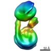
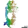
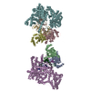
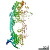
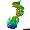
| Title | Structure of the 4_601_157 tetranucleosome (C2 form) | ||||||||||||
 Components Components |
| ||||||||||||
 Keywords Keywords | GENE REGULATION / nucleosome / histone / DNA / tetranucleosome / chromatin fiber | ||||||||||||
| Function / homology |  Function and homology information Function and homology informationstructural constituent of chromatin / nucleosome / nucleosome assembly / heterochromatin formation / protein heterodimerization activity / DNA binding / nucleoplasm / nucleus Similarity search - Function | ||||||||||||
| Biological species | synthetic construct (others) | ||||||||||||
| Method |  X-RAY DIFFRACTION / SYNCHROTRON / MOLECULAR REPLACEMENT / Resolution: 6.721 Å X-RAY DIFFRACTION / SYNCHROTRON / MOLECULAR REPLACEMENT / Resolution: 6.721 Å | ||||||||||||
 Authors Authors | Ekundayo, B. / Schalch, T. | ||||||||||||
| Funding support |  Switzerland, 3items Switzerland, 3items
| ||||||||||||
 Citation Citation | Journal: J. Mol. Biol. / Year: 2017 Title: Capturing Structural Heterogeneity in Chromatin Fibers. Authors: Ekundayo, B. / Richmond, T.J. / Schalch, T. | ||||||||||||
| History |
|
- Structure visualization
| Structure viewer | Molecule: MolmilJmol/JSmol |
|---|
- Downloads & links
-Download
| PDBx/mmCIF format | 5oxv.cif.gz | 611.9 KB | Display | PDBx/mmCIF format |
|---|---|---|---|---|
| PDB format | pdb5oxv.ent.gz | 467.5 KB | Display | PDB format |
| PDBx/mmJSON format | 5oxv.json.gz | Tree view | PDBx/mmJSON format | |
| Others |  Other downloads Other downloads |
-Validation report
| Arichive directory | https://data.pdbj.org/pub/pdb/validation_reports/ox/5oxvftp://data.pdbj.org/pub/pdb/validation_reports/ox/5oxv | HTTPS FTP |
|---|
-Related structure data
| Related structure data | 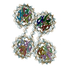 5oy7C  3lz0S C: citing same article ( S: Starting model for refinement |
|---|---|
| Similar structure data |


-Links
 PDBj
PDBj


- Assembly
| Deposited unit | 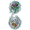
| ||||||||
|---|---|---|---|---|---|---|---|---|---|
| 1 |
| ||||||||
| Unit cell |
|
-Components
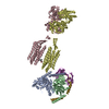
-DNA chain , 2 types, 2 molecules JI
| #1: DNA chain | Mass: 96886.500 Da / Num. of mol.: 1 Source method: isolated from a genetically manipulated source Source: (gene. exp.) synthetic construct (others) / Production host:  |
|---|---|
| #6: DNA chain | Mass: 96147.062 Da / Num. of mol.: 1 Source method: isolated from a genetically manipulated source Source: (gene. exp.) synthetic construct (others) / Production host: |
-Protein , 4 types, 16 molecules RNHDQMGCPLFBOKEA
| #2: Protein | Mass: 13979.291 Da / Num. of mol.: 4 Source method: isolated from a genetically manipulated source Source: (gene. exp.) #3: Protein | Mass: 14109.436 Da / Num. of mol.: 4 Source method: isolated from a genetically manipulated source Source: (gene. exp.) #4: Protein | Mass: 11263.231 Da / Num. of mol.: 4 Source method: isolated from a genetically manipulated source Source: (gene. exp.) #5: Protein | Mass: 15303.930 Da / Num. of mol.: 4 Source method: isolated from a genetically manipulated source Source: (gene. exp.) |
|---|
-Experimental details
-Experiment
| Experiment | Method: X-RAY DIFFRACTION / Number of used crystals: 1 |
|---|
- Sample preparation
| Crystal | Density Matthews: 2.99 Å3/Da / Density % sol: 58.87 % |
|---|---|
| Crystal grow | Temperature: 291 K / Method: vapor diffusion / pH: 6 Details: 30-60 mM KCl, 90-110 mM MgCl2 and 5 mM Na-cacodylate, pH 6 |
-Data collection
| Diffraction | Mean temperature: 100 K |
|---|---|
| Diffraction source | Source: SYNCHROTRON / Site: SLS / Beamline: X06DA / Wavelength: 1 Å |
| Detector | Type: DECTRIS PILATUS 2M-F / Detector: PIXEL / Date: Oct 11, 2014 |
| Radiation | Protocol: SINGLE WAVELENGTH / Monochromatic (M) / Laue (L): M / Scattering type: x-ray |
| Radiation wavelength | Wavelength: 1 Å / Relative weight: 1 |
| Reflection | Resolution: 6.72→200 Å / Num. obs: 9194 / % possible obs: 100 % / Redundancy: 6.5 % / CC1/2: 0.98 / Rmerge(I) obs: 0.163 / Net I/σ(I): 5.8 |
| Reflection shell | Resolution: 6.72→7.51 Å / Redundancy: 7.4 % / Rmerge(I) obs: 4.943 / Mean I/σ(I) obs: 0.3 / Num. unique obs: 2554 / CC1/2: 0.164 / % possible all: 100 |
- Processing
| Software |
| ||||||||||||||||||||||||||||||||||||||||||||||||||||||||
|---|---|---|---|---|---|---|---|---|---|---|---|---|---|---|---|---|---|---|---|---|---|---|---|---|---|---|---|---|---|---|---|---|---|---|---|---|---|---|---|---|---|---|---|---|---|---|---|---|---|---|---|---|---|---|---|---|---|
| Refinement | Method to determine structure: MOLECULAR REPLACEMENT Starting model: 3LZ0 Resolution: 6.721→71.666 Å / SU ML: 1.49 / Cross valid method: FREE R-VALUE / σ(F): 1.33 / Phase error: 58.89
| ||||||||||||||||||||||||||||||||||||||||||||||||||||||||
| Solvent computation | Shrinkage radii: 0.9 Å / VDW probe radii: 1.11 Å | ||||||||||||||||||||||||||||||||||||||||||||||||||||||||
| Refinement step | Cycle: LAST / Resolution: 6.721→71.666 Å
| ||||||||||||||||||||||||||||||||||||||||||||||||||||||||
| Refine LS restraints |
| ||||||||||||||||||||||||||||||||||||||||||||||||||||||||
| LS refinement shell |
|
