 Movie
Movie Controller

[English] 日本語
 Yorodumi
- PDB-3fyt: Crystal structure of Bacillus pumilus acetyl xylan esterase S181A... -
+ Open data

- Basic information
| Entry | Database: PDB / ID: 3fyt | |||||||||
|---|---|---|---|---|---|---|---|---|---|---|
| Title | Crystal structure of Bacillus pumilus acetyl xylan esterase S181A mutant in complex with beta-D-xylopyranose | |||||||||
 Components Components | Acetyl xylan esterase | |||||||||
 Keywords Keywords | HYDROLASE / alpha/beta hydrolase / carbohydrate esterase / CE7 / Bacillus pumilus | |||||||||
| Function / homology |  Function and homology information Function and homology informationacetylesterase / polysaccharide metabolic process / acetylesterase activity Similarity search - Function | |||||||||
| Biological species |  | |||||||||
| Method |  X-RAY DIFFRACTION / SYNCHROTRON / FOURIER SYNTHESIS / molecular replacement / Resolution: 2.58 Å X-RAY DIFFRACTION / SYNCHROTRON / FOURIER SYNTHESIS / molecular replacement / Resolution: 2.58 Å | |||||||||
 Authors Authors | Krastanova, I. / Cassetta, A. / Lamba, D. | |||||||||
 Citation Citation | Journal: To be Published Title: Structural and functional studies of Bacillus pumilus acetyl xylan esterase Authors: Krastanova, I. / Cassetta, A. / Mastihubova, M. / Biely, P. / Lamba, D. #1: Journal: Biochim.Biophys.Acta / Year: 2005Title: Heterologous expression, purification, crystallization, X-ray analysis and phasing of the acetyl xylan esterase from Bacillus pumilus Authors: Krastanova, I. / Guarnaccia, C. / Zahariev, S. / Degrassi, G. / Lamba, D. #2: Journal: Microbiology / Year: 2000Title: The acetyl xylan esterase of Bacillus pumilus belongs to a family of esterases with broad substrate specificity Authors: Degrassi, G. / Kojic, M. / Ljubijankic, G. / Venturi, V. #3: Journal: Appl.Environ.Microbiol. / Year: 1998Title: Purification and characterization of an acetyl xylan esterase from Bacillus pumilus Authors: Degrassi, G. / Okeke, B.C. / Bruschi, C.V. / Venturi, V. | |||||||||
| History |
|
- Structure visualization
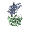
| Structure viewer | Molecule: MolmilJmol/JSmol |
|---|
- Downloads & links
-Download
| PDBx/mmCIF format | 3fyt.cif.gz | 727.3 KB | Display | PDBx/mmCIF format |
|---|---|---|---|---|
| PDB format | pdb3fyt.ent.gz | 607 KB | Display | PDB format |
| PDBx/mmJSON format | 3fyt.json.gz | Tree view | PDBx/mmJSON format | |
| Others |  Other downloads Other downloads |
-Validation report
| Arichive directory | https://data.pdbj.org/pub/pdb/validation_reports/fy/3fytftp://data.pdbj.org/pub/pdb/validation_reports/fy/3fyt | HTTPS FTP |
|---|
-Related structure data
| Related structure data |  3fvrSC  3fvtC 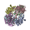 3fyuC S: Starting model for refinement C: citing same article ( |
|---|---|
| Similar structure data |


-Links
 PDBj
PDBj- Assembly
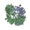
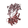
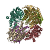
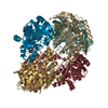
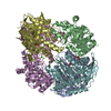
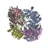
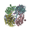
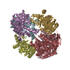
| Deposited unit | 
| ||||||||
|---|---|---|---|---|---|---|---|---|---|
| 1 | 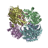
| ||||||||
| 2 | 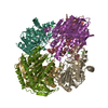
| ||||||||
| Unit cell |
|
-Components
| #1: Protein | Mass: 36098.844 Da / Num. of mol.: 12 / Mutation: S181A Source method: isolated from a genetically manipulated source Source: (gene. exp.) #2: Chemical | ChemComp-CL /   Mass: 35.453 Da / Num. of mol.: 34 / Source method: obtained synthetically / Formula: Cl Mass: 35.453 Da / Num. of mol.: 34 / Source method: obtained synthetically / Formula: Cl#3: Sugar | ChemComp-XYP / 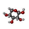  Type: D-saccharide, beta linking / Mass: 150.130 Da / Num. of mol.: 6 Type: D-saccharide, beta linking / Mass: 150.130 Da / Num. of mol.: 6Source method: isolated from a genetically manipulated source Formula: C5H10O5 #4: Water | ChemComp-HOH / |  Mass: 18.015 Da / Num. of mol.: 362 / Source method: isolated from a natural source / Formula: H2O Mass: 18.015 Da / Num. of mol.: 362 / Source method: isolated from a natural source / Formula: H2O |
|---|
-Experimental details
-Experiment
| Experiment | Method: X-RAY DIFFRACTION / Number of used crystals: 1 |
|---|
- Sample preparation
| Crystal | Density Matthews: 2.44 Å3/Da / Density % sol: 49.63 % |
|---|---|
| Crystal grow | Temperature: 293 K / Method: microbatch / pH: 6 Details: 0.20M lithium chloride, 0.13M D-xylose, 3% PEG 6000, 0.03M MES, pH 6.0, microbatch, temperature 293K |
-Data collection
| Diffraction | Mean temperature: 100 K |
|---|---|
| Diffraction source | Source: SYNCHROTRON / Site: ESRF  / Beamline: ID14-4 / Wavelength: 0.978 Å / Beamline: ID14-4 / Wavelength: 0.978 Å |
| Detector | Type: ADSC QUANTUM 4 / Detector: CCD / Date: Sep 16, 2005 |
| Radiation | Monochromator: Si (111) / Protocol: SINGLE WAVELENGTH / Monochromatic (M) / Laue (L): M / Scattering type: x-ray |
| Radiation wavelength | Wavelength: 0.978 Å / Relative weight: 1 |
| Reflection | Resolution: 2.58→24.83 Å / Num. all: 100895 / Num. obs: 100895 / % possible obs: 77.8 % / Observed criterion σ(F): 0 / Observed criterion σ(I): -3 / Redundancy: 3.8 % / Biso Wilson estimate: 37.66 Å2 / Rmerge(I) obs: 0.064 / Χ2: 1.117 |
| Reflection shell | Resolution: 2.58→2.66 Å / Redundancy: 3 % / Rmerge(I) obs: 0.18 / Mean I/σ(I) obs: 4 / Num. unique all: 4985 / Χ2: 1.021 / % possible all: 50.1 |
-Phasing
| Phasing | Method: molecular replacement |
|---|
- Processing
| Software |
| ||||||||||||||||||||||||||||||||
|---|---|---|---|---|---|---|---|---|---|---|---|---|---|---|---|---|---|---|---|---|---|---|---|---|---|---|---|---|---|---|---|---|---|
| Refinement | Method to determine structure: FOURIER SYNTHESIS Starting model: PDB entry 3FVR Resolution: 2.58→24.83 Å / Occupancy max: 1 / Occupancy min: 1 / Cross valid method: THROUGHOUT / σ(F): 0 / Stereochemistry target values: Engh & Huber
| ||||||||||||||||||||||||||||||||
| Solvent computation | Bsol: 39.298 Å2 | ||||||||||||||||||||||||||||||||
| Displacement parameters | Biso max: 100 Å2 / Biso mean: 28.857 Å2 / Biso min: 5 Å2
| ||||||||||||||||||||||||||||||||
| Refinement step | Cycle: LAST / Resolution: 2.58→24.83 Å
| ||||||||||||||||||||||||||||||||
| Refine LS restraints |
| ||||||||||||||||||||||||||||||||
| Xplor file |
|
