 Movie
Movie Controller

[English] 日本語
 Yorodumi
- PDB-7e6o: Crystal structure of polyol dehydrogenase from Paracoccus denitri... -
+ Open data

- Basic information
| Entry | Database: PDB / ID: 7e6o | ||||||
|---|---|---|---|---|---|---|---|
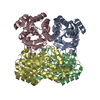
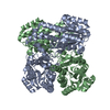
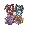
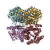
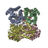
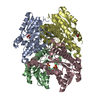
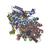
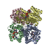
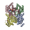
| Title | Crystal structure of polyol dehydrogenase from Paracoccus denitrificans | ||||||
 Components Components | Short-chain dehydrogenase/reductase SDR | ||||||
 Keywords Keywords | OXIDOREDUCTASE / short-chain dehydrogenase / sorbitol oxidation | ||||||
| Function / homology | short chain dehydrogenase / PKS_KR / Short-chain dehydrogenase/reductase, conserved site / Short-chain dehydrogenases/reductases family signature. / Short-chain dehydrogenase/reductase SDR / oxidoreductase activity / NAD(P)-binding domain superfamily / Short-chain dehydrogenase/reductase SDR Function and homology information Function and homology information | ||||||
| Biological species |  Paracoccus denitrificans (bacteria) Paracoccus denitrificans (bacteria) | ||||||
| Method |  X-RAY DIFFRACTION / SYNCHROTRON / MOLECULAR REPLACEMENT / Resolution: 2.1 Å X-RAY DIFFRACTION / SYNCHROTRON / MOLECULAR REPLACEMENT / Resolution: 2.1 Å | ||||||
 Authors Authors | Sha, F. / Zheng, Y.C. | ||||||
 Citation Citation | Journal: To Be Published Title: Crystal structure of polyol dehydrogenase from Paracoccus denitrificans Authors: Sha, F. / Zheng, Y.C. / Gao, J. | ||||||
| History |
|
- Structure visualization
| Structure viewer | Molecule: MolmilJmol/JSmol |
|---|
- Downloads & links
-Download
| PDBx/mmCIF format | 7e6o.cif.gz | 197.6 KB | Display | PDBx/mmCIF format |
|---|---|---|---|---|
| PDB format | pdb7e6o.ent.gz | 156.3 KB | Display | PDB format |
| PDBx/mmJSON format | 7e6o.json.gz | Tree view | PDBx/mmJSON format | |
| Others |  Other downloads Other downloads |
-Validation report
| Arichive directory | https://data.pdbj.org/pub/pdb/validation_reports/e6/7e6oftp://data.pdbj.org/pub/pdb/validation_reports/e6/7e6o | HTTPS FTP |
|---|
-Related structure data
| Related structure data | 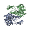 1k2wS S: Starting model for refinement |
|---|---|
| Similar structure data |

-Links
 PDBj
PDBj


- Assembly
| Deposited unit | 
| ||||||||
|---|---|---|---|---|---|---|---|---|---|
| 1 |
| ||||||||
| Unit cell |
|
-Components
| #1: Protein | Mass: 27780.877 Da / Num. of mol.: 4 / Mutation: None Source method: isolated from a genetically manipulated source Source: (gene. exp.) Paracoccus denitrificans (strain Pd 1222) (bacteria)Strain: Pd 1222 / Gene: Pden_4841 / Production host: #2: Water | ChemComp-HOH / |  Mass: 18.015 Da / Num. of mol.: 503 / Source method: isolated from a natural source / Formula: H2O Mass: 18.015 Da / Num. of mol.: 503 / Source method: isolated from a natural source / Formula: H2O |
|---|
-Experimental details
-Experiment
| Experiment | Method: X-RAY DIFFRACTION / Number of used crystals: 1 |
|---|
- Sample preparation
| Crystal | Density Matthews: 2.05 Å3/Da / Density % sol: 39.94 % |
|---|---|
| Crystal grow | Temperature: 291 K / Method: evaporation / pH: 6.5 / Details: 0.1 M MES, pH 6.5 15% (w/v) PEG 6000 5% (w/v) MPD |
-Data collection
| Diffraction | Mean temperature: 100 K / Serial crystal experiment: N |
|---|---|
| Diffraction source | Source: SYNCHROTRON / Site: SSRF  / Beamline: BL17U1 / Wavelength: 0.9789 Å / Beamline: BL17U1 / Wavelength: 0.9789 Å |
| Detector | Type: ADSC QUANTUM 315r / Detector: CCD / Date: Jun 18, 2019 |
| Radiation | Protocol: SINGLE WAVELENGTH / Monochromatic (M) / Laue (L): M / Scattering type: x-ray |
| Radiation wavelength | Wavelength: 0.9789 Å / Relative weight: 1 |
| Reflection | Resolution: 2.1→50 Å / Num. obs: 686828 / % possible obs: 99.4 % / Redundancy: 12.8 % / CC1/2: 0.991 / Net I/σ(I): 20.37 |
| Reflection shell | Resolution: 2.1→2.14 Å / Mean I/σ(I) obs: 2.9 / Num. unique obs: 2623 / CC1/2: 0.775 / CC star: 0.934 / Χ2: 0.939 |
- Processing
| Software |
| ||||||||||||||||||||||||||||||||||||||||||||||||||||||||||||
|---|---|---|---|---|---|---|---|---|---|---|---|---|---|---|---|---|---|---|---|---|---|---|---|---|---|---|---|---|---|---|---|---|---|---|---|---|---|---|---|---|---|---|---|---|---|---|---|---|---|---|---|---|---|---|---|---|---|---|---|---|---|
| Refinement | Method to determine structure: MOLECULAR REPLACEMENT Starting model: 1K2W Resolution: 2.1→43.97 Å / Cor.coef. Fo:Fc: 0.96 / Cor.coef. Fo:Fc free: 0.938 / SU B: 4.611 / SU ML: 0.12 / Cross valid method: THROUGHOUT / σ(F): 0 / ESU R: 0.231 / ESU R Free: 0.179 / Stereochemistry target values: MAXIMUM LIKELIHOOD Details: HYDROGENS HAVE BEEN ADDED IN THE RIDING POSITIONS U VALUES : REFINED INDIVIDUALLY
| ||||||||||||||||||||||||||||||||||||||||||||||||||||||||||||
| Solvent computation | Ion probe radii: 0.8 Å / Shrinkage radii: 0.8 Å / VDW probe radii: 1.2 Å / Solvent model: MASK | ||||||||||||||||||||||||||||||||||||||||||||||||||||||||||||
| Displacement parameters | Biso max: 138.03 Å2 / Biso mean: 28.542 Å2 / Biso min: 15.21 Å2
| ||||||||||||||||||||||||||||||||||||||||||||||||||||||||||||
| Refinement step | Cycle: final / Resolution: 2.1→43.97 Å
| ||||||||||||||||||||||||||||||||||||||||||||||||||||||||||||
| Refine LS restraints |
| ||||||||||||||||||||||||||||||||||||||||||||||||||||||||||||
| LS refinement shell | Resolution: 2.104→2.159 Å / Rfactor Rfree error: 0 / Total num. of bins used: 20
|
