 Movie
Movie Controller


+ Open data

- Basic information
| Entry | Database: PDB / ID: 1k2w | ||||||
|---|---|---|---|---|---|---|---|
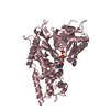
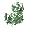
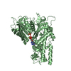
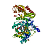
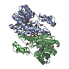
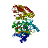
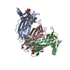
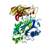
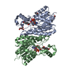
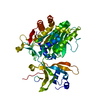
| Title | Crystal structure of sorbitol dehydrogenase from R. sphaeroides | ||||||
 Components Components | SORBITOL DEHYDROGENASE | ||||||
 Keywords Keywords | OXIDOREDUCTASE / short-chain dehydrogenase / enzyme | ||||||
| Function / homology |  Function and homology information Function and homology informationgalactitol 2-dehydrogenase / galactitol 2-dehydrogenase activity / L-iditol 2-dehydrogenase / L-iditol 2-dehydrogenase (NAD+) activity / Oxidoreductases; Acting on the CH-OH group of donors; With NAD+ or NADP+ as acceptor Similarity search - Function | ||||||
| Biological species |  Rhodobacter sphaeroides (bacteria) Rhodobacter sphaeroides (bacteria) | ||||||
| Method |  X-RAY DIFFRACTION / MOLECULAR REPLACEMENT / Resolution: 2.4 Å X-RAY DIFFRACTION / MOLECULAR REPLACEMENT / Resolution: 2.4 Å | ||||||
 Authors Authors | Philippsen, A. / Schirmer, T. / Stetefeld, J. | ||||||
 Citation Citation | Journal: Acta Crystallogr.,Sect.D / Year: 2005 Title: Structure of zinc-independent sorbitol dehydrogenase from Rhodobacter sphaeroides at 2.4 A resolution. Authors: Philippsen, A. / Schirmer, T. / Stein, M.A. / Giffhorn, F. / Stetefeld, J. #1: Journal: Microbiology (Reading, Engl.) / Year: 1995Title: Polyol metabolism of Rhodobacter sphaeroides: biochemical characterization of a short-chain sorbitol dehydrogenase Authors: Schauder, S. / Schneider, K.H. / Giffhorn, F. | ||||||
| History |
|
- Structure visualization
| Structure viewer | Molecule: MolmilJmol/JSmol |
|---|
- Downloads & links
-Download
| PDBx/mmCIF format | 1k2w.cif.gz | 107.4 KB | Display | PDBx/mmCIF format |
|---|---|---|---|---|
| PDB format | pdb1k2w.ent.gz | 83.5 KB | Display | PDB format |
| PDBx/mmJSON format | 1k2w.json.gz | Tree view | PDBx/mmJSON format | |
| Others |  Other downloads Other downloads |
-Validation report
| Arichive directory | https://data.pdbj.org/pub/pdb/validation_reports/k2/1k2wftp://data.pdbj.org/pub/pdb/validation_reports/k2/1k2w | HTTPS FTP |
|---|
-Related structure data
| Related structure data |  1gegS S: Starting model for refinement |
|---|---|
| Similar structure data |
-Links
 PDBj
PDBj


- Assembly
| Deposited unit | 
| ||||||||
|---|---|---|---|---|---|---|---|---|---|
| 1 |
| ||||||||
| 2 | 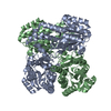
| ||||||||
| 3 | 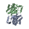
| ||||||||
| Unit cell |
| ||||||||
| Details | biological assembly is dimer according to sedimentation analysis |
-Components
| #1: Protein | Mass: 27040.840 Da / Num. of mol.: 2 Source method: isolated from a genetically manipulated source Source: (gene. exp.) Rhodobacter sphaeroides (bacteria) / Production host: #2: Water | ChemComp-HOH / |  Mass: 18.015 Da / Num. of mol.: 170 / Source method: isolated from a natural source / Formula: H2O Mass: 18.015 Da / Num. of mol.: 170 / Source method: isolated from a natural source / Formula: H2O |
|---|
-Experimental details
-Experiment
| Experiment | Method: X-RAY DIFFRACTION / Number of used crystals: 1 |
|---|
- Sample preparation
| Crystal | Density Matthews: 2.29 Å3/Da / Density % sol: 45.91 % | ||||||||||||||||||||||||||||||
|---|---|---|---|---|---|---|---|---|---|---|---|---|---|---|---|---|---|---|---|---|---|---|---|---|---|---|---|---|---|---|---|
| Crystal grow | Temperature: 300 K / Method: vapor diffusion, hanging drop / pH: 7 Details: PEG4000, methanol, pH 7.0, VAPOR DIFFUSION, HANGING DROP, temperature 300K | ||||||||||||||||||||||||||||||
| Crystal grow | *PLUS Method: vapor diffusion, sitting drop | ||||||||||||||||||||||||||||||
| Components of the solutions | *PLUS
|
-Data collection
| Diffraction | Mean temperature: 280 K |
|---|---|
| Diffraction source | Source: ROTATING ANODE / Type: OTHER / Wavelength: 1.5418 |
| Detector | Type: MARRESEARCH / Detector: IMAGE PLATE / Date: Jan 1, 1999 |
| Radiation | Protocol: SINGLE WAVELENGTH / Monochromatic (M) / Laue (L): M / Scattering type: x-ray |
| Radiation wavelength | Wavelength: 1.5418 Å / Relative weight: 1 |
| Reflection | Resolution: 2.3→30 Å / Num. all: 22993 / Num. obs: 21038 / % possible obs: 91.5 % / Observed criterion σ(F): 2.5 / Observed criterion σ(I): 2.5 / Redundancy: 3 % / Biso Wilson estimate: 25.7 Å2 / Rmerge(I) obs: 0.094 / Rsym value: 0.079 / Net I/σ(I): 8 |
| Reflection shell | Resolution: 2.3→2.42 Å / % possible all: 73.3 |
| Reflection | *PLUS Highest resolution: 2.4 Å / Lowest resolution: 30 Å / Rmerge(I) obs: 0.078 |
| Reflection shell | *PLUS Highest resolution: 2.42 Å / Lowest resolution: 2.57 Å / % possible obs: 92.1 % / Redundancy: 2.4 % / Rmerge(I) obs: 0.225 / Mean I/σ(I) obs: 3.2 |
- Processing
| Software |
| |||||||||||||||||||||||||
|---|---|---|---|---|---|---|---|---|---|---|---|---|---|---|---|---|---|---|---|---|---|---|---|---|---|---|
| Refinement | Method to determine structure: MOLECULAR REPLACEMENT Starting model: PDB Entry 1GEG Resolution: 2.4→30 Å / Cross valid method: THROUGHOUT / Stereochemistry target values: Engh & Huber
| |||||||||||||||||||||||||
| Refinement step | Cycle: LAST / Resolution: 2.4→30 Å
| |||||||||||||||||||||||||
| Refine LS restraints |
| |||||||||||||||||||||||||
| Refinement | *PLUS Lowest resolution: 30 Å | |||||||||||||||||||||||||
| Solvent computation | *PLUS | |||||||||||||||||||||||||
| Displacement parameters | *PLUS | |||||||||||||||||||||||||
| Refine LS restraints | *PLUS
|
