 Movie
Movie Controller

[English] 日本語
 Yorodumi
- PDB-5f9c: Crystal structure of the G121R mutant of human phosphoglucomutase 1 -
+ Open data

- Basic information
| Entry | Database: PDB / ID: 5f9c | ||||||
|---|---|---|---|---|---|---|---|
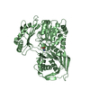
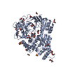
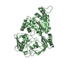
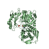
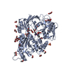
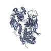
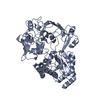
| Title | Crystal structure of the G121R mutant of human phosphoglucomutase 1 | ||||||
 Components Components | Phosphoglucomutase-1 | ||||||
 Keywords Keywords | ISOMERASE / isomerase metabolism | ||||||
| Function / homology |  Function and homology information Function and homology informationDefective PGM1 causes PGM1-CDG / phosphoglucomutase (alpha-D-glucose-1,6-bisphosphate-dependent) / phosphoglucomutase activity / beta-D-galactose catabolic process via UDP-galactose, Leloir pathway / Galactose catabolism / glycogen catabolic process / Glycogen breakdown (glycogenolysis) / Glycogen synthesis / glycolytic process / gluconeogenesis ...Defective PGM1 causes PGM1-CDG / phosphoglucomutase (alpha-D-glucose-1,6-bisphosphate-dependent) / phosphoglucomutase activity / beta-D-galactose catabolic process via UDP-galactose, Leloir pathway / Galactose catabolism / glycogen catabolic process / Glycogen breakdown (glycogenolysis) / Glycogen synthesis / glycolytic process / gluconeogenesis / glucose metabolic process / tertiary granule lumen / carbohydrate metabolic process / ficolin-1-rich granule lumen / Neutrophil degranulation / magnesium ion binding / extracellular exosome / extracellular region / cytoplasm / cytosol Similarity search - Function | ||||||
| Biological species |  Homo sapiens (human) Homo sapiens (human) | ||||||
| Method |  X-RAY DIFFRACTION / SYNCHROTRON / Resolution: 2.5 Å X-RAY DIFFRACTION / SYNCHROTRON / Resolution: 2.5 Å | ||||||
 Authors Authors | Beamer, L.J. | ||||||
 Citation Citation | Journal: J.Mol.Biol. / Year: 2016 Title: Induced Structural Disorder as a Molecular Mechanism for Enzyme Dysfunction in Phosphoglucomutase 1 Deficiency. Authors: Stiers, K.M. / Kain, B.N. / Graham, A.C. / Beamer, L.J. | ||||||
| History |
|
- Structure visualization
| Structure viewer | Molecule: MolmilJmol/JSmol |
|---|
- Downloads & links
-Download
| PDBx/mmCIF format | 5f9c.cif.gz | 426.1 KB | Display | PDBx/mmCIF format |
|---|---|---|---|---|
| PDB format | pdb5f9c.ent.gz | 351.5 KB | Display | PDB format |
| PDBx/mmJSON format | 5f9c.json.gz | Tree view | PDBx/mmJSON format | |
| Others |  Other downloads Other downloads |
-Validation report
| Arichive directory | https://data.pdbj.org/pub/pdb/validation_reports/f9/5f9cftp://data.pdbj.org/pub/pdb/validation_reports/f9/5f9c | HTTPS FTP |
|---|
-Related structure data
| Related structure data |  5epcSC  5hshC S: Starting model for refinement C: citing same article ( |
|---|---|
| Similar structure data |

-Links
 PDBj
PDBj


- Assembly
| Deposited unit | 
| ||||||||
|---|---|---|---|---|---|---|---|---|---|
| 1 | 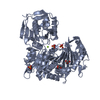
| ||||||||
| 2 | 
| ||||||||
| Unit cell |
|
-Components
| #1: Protein | Mass: 61619.027 Da / Num. of mol.: 2 / Mutation: G121R Source method: isolated from a genetically manipulated source Source: (gene. exp.) Homo sapiens (human) / Gene: PGM1 / Production host:  References: UniProt: P36871, phosphoglucomutase (alpha-D-glucose-1,6-bisphosphate-dependent) #2: Chemical |   Mass: 24.305 Da / Num. of mol.: 2 / Source method: obtained synthetically / Formula: Mg Mass: 24.305 Da / Num. of mol.: 2 / Source method: obtained synthetically / Formula: Mg#3: Chemical | ChemComp-SO4 /   Mass: 96.063 Da / Num. of mol.: 9 / Source method: obtained synthetically / Formula: SO4 Mass: 96.063 Da / Num. of mol.: 9 / Source method: obtained synthetically / Formula: SO4#4: Chemical |   Mass: 92.094 Da / Num. of mol.: 2 / Source method: obtained synthetically / Formula: C3H8O3 Mass: 92.094 Da / Num. of mol.: 2 / Source method: obtained synthetically / Formula: C3H8O3#5: Water | ChemComp-HOH / |  Mass: 18.015 Da / Num. of mol.: 80 / Source method: isolated from a natural source / Formula: H2O Mass: 18.015 Da / Num. of mol.: 80 / Source method: isolated from a natural source / Formula: H2O |
|---|
-Experimental details
-Experiment
| Experiment | Method: X-RAY DIFFRACTION / Number of used crystals: 1 |
|---|
- Sample preparation
| Crystal | Density Matthews: 2.97 Å3/Da / Density % sol: 58.65 % |
|---|---|
| Crystal grow | Temperature: 293 K / Method: vapor diffusion, hanging drop Details: 1.5 M ammonium sulfate with 0.15 lithium sulfate and 0.1 CAPS buffer, pH 10.5 PH range: 10.5 |
-Data collection
| Diffraction | Mean temperature: 100 K |
|---|---|
| Diffraction source | Source: SYNCHROTRON / Site: ALS  / Beamline: 4.2.2 / Wavelength: 1.00001 Å / Beamline: 4.2.2 / Wavelength: 1.00001 Å |
| Detector | Type: RDI CMOS_8M / Detector: CMOS / Date: Oct 25, 2015 |
| Radiation | Protocol: SINGLE WAVELENGTH / Monochromatic (M) / Laue (L): M / Scattering type: x-ray |
| Radiation wavelength | Wavelength: 1.00001 Å / Relative weight: 1 |
| Reflection | Resolution: 2.35→60.943 Å / Num. obs: 71761 / % possible obs: 99.8 % / Redundancy: 14.1 % / Biso Wilson estimate: 55.3 Å2 / Rmerge(I) obs: 0.123 / Net I/σ(I): 16.5 |
| Reflection shell | Resolution: 2.35→2.41 Å / Redundancy: 12.4 % / Rmerge(I) obs: 3.101 / Mean I/σ(I) obs: 0.8 / % possible all: 99.2 |
- Processing
| Software |
| |||||||||||||||||||||||||||||||||||||||||||||||||||||||||||||||||||||||||||||||||||||||||||||||||||||||||||||||||||||||||||||||||||||||||||||||||||||||||||||||||||||||||||||||||||||||||||||||||||||||||||||||||||||||||
|---|---|---|---|---|---|---|---|---|---|---|---|---|---|---|---|---|---|---|---|---|---|---|---|---|---|---|---|---|---|---|---|---|---|---|---|---|---|---|---|---|---|---|---|---|---|---|---|---|---|---|---|---|---|---|---|---|---|---|---|---|---|---|---|---|---|---|---|---|---|---|---|---|---|---|---|---|---|---|---|---|---|---|---|---|---|---|---|---|---|---|---|---|---|---|---|---|---|---|---|---|---|---|---|---|---|---|---|---|---|---|---|---|---|---|---|---|---|---|---|---|---|---|---|---|---|---|---|---|---|---|---|---|---|---|---|---|---|---|---|---|---|---|---|---|---|---|---|---|---|---|---|---|---|---|---|---|---|---|---|---|---|---|---|---|---|---|---|---|---|---|---|---|---|---|---|---|---|---|---|---|---|---|---|---|---|---|---|---|---|---|---|---|---|---|---|---|---|---|---|---|---|---|---|---|---|---|---|---|---|---|---|---|---|---|---|---|---|---|
| Refinement | Starting model: 5EPC Resolution: 2.5→60.943 Å / SU ML: 0.35 / Cross valid method: FREE R-VALUE / σ(F): 1.37 / Phase error: 28.33 / Stereochemistry target values: ML
| |||||||||||||||||||||||||||||||||||||||||||||||||||||||||||||||||||||||||||||||||||||||||||||||||||||||||||||||||||||||||||||||||||||||||||||||||||||||||||||||||||||||||||||||||||||||||||||||||||||||||||||||||||||||||
| Solvent computation | Shrinkage radii: 0.9 Å / VDW probe radii: 1.11 Å / Solvent model: FLAT BULK SOLVENT MODEL | |||||||||||||||||||||||||||||||||||||||||||||||||||||||||||||||||||||||||||||||||||||||||||||||||||||||||||||||||||||||||||||||||||||||||||||||||||||||||||||||||||||||||||||||||||||||||||||||||||||||||||||||||||||||||
| Refinement step | Cycle: LAST / Resolution: 2.5→60.943 Å
| |||||||||||||||||||||||||||||||||||||||||||||||||||||||||||||||||||||||||||||||||||||||||||||||||||||||||||||||||||||||||||||||||||||||||||||||||||||||||||||||||||||||||||||||||||||||||||||||||||||||||||||||||||||||||
| Refine LS restraints |
| |||||||||||||||||||||||||||||||||||||||||||||||||||||||||||||||||||||||||||||||||||||||||||||||||||||||||||||||||||||||||||||||||||||||||||||||||||||||||||||||||||||||||||||||||||||||||||||||||||||||||||||||||||||||||
| LS refinement shell |
| |||||||||||||||||||||||||||||||||||||||||||||||||||||||||||||||||||||||||||||||||||||||||||||||||||||||||||||||||||||||||||||||||||||||||||||||||||||||||||||||||||||||||||||||||||||||||||||||||||||||||||||||||||||||||
| Refinement TLS params. | Method: refined / Refine-ID: X-RAY DIFFRACTION
| |||||||||||||||||||||||||||||||||||||||||||||||||||||||||||||||||||||||||||||||||||||||||||||||||||||||||||||||||||||||||||||||||||||||||||||||||||||||||||||||||||||||||||||||||||||||||||||||||||||||||||||||||||||||||
| Refinement TLS group |
|
