Isoform 2 of Nuclear distribution protein nudE homolog 1
Ras-related protein Rab-9A
Keywords
PROTEIN TRANSPORT / Rab9A / Nde1 / Rab GTPase / effector
Function / homology
Function and homology information
: / Rab protein signal transduction / Retrograde transport at the Trans-Golgi-Network / RAB geranylgeranylation / RHOBTB3 ATPase cycle / spindle pole centrosome / microtubule nucleation / RAB GEFs exchange GTP for GDP on RABs / mitotic centrosome separation / vesicle transport along microtubule ...: / Rab protein signal transduction / Retrograde transport at the Trans-Golgi-Network / RAB geranylgeranylation / RHOBTB3 ATPase cycle / spindle pole centrosome / microtubule nucleation / RAB GEFs exchange GTP for GDP on RABs / mitotic centrosome separation / vesicle transport along microtubule / retrograde transport, endosome to Golgi / kinesin complex / centrosome localization / cleavage furrow / centrosome duplication / establishment of mitotic spindle orientation / positive regulation of exocytosis / neuroblast proliferation / transport vesicle / phagocytic vesicle / Amplification of signal from unattached kinetochores via a MAD2 inhibitory signal / Loss of Nlp from mitotic centrosomes / Loss of proteins required for interphase microtubule organization from the centrosome / Recruitment of mitotic centrosome proteins and complexes / receptor-mediated endocytosis / Mitotic Prometaphase / Recruitment of NuMA to mitotic centrosomes / Anchoring of the basal body to the plasma membrane / EML4 and NUDC in mitotic spindle formation / AURKA Activation by TPX2 / Resolution of Sister Chromatid Cohesion / trans-Golgi network membrane / small monomeric GTPase / chromosome segregation / RHO GTPases Activate Formins / cerebral cortex development / kinetochore / phagocytic vesicle membrane / neuron migration / GDP binding / Separation of Sister Chromatids / melanosome / Regulation of PLK1 Activity at G2/M Transition / late endosome / cell migration / protein transport / regulation of protein localization / G protein activity / microtubule binding / microtubule / lysosome / Golgi membrane / cell division / GTPase activity / synapse / centrosome / endoplasmic reticulum membrane / GTP binding / extracellular exosome / identical protein binding / membrane / plasma membrane / cytosol Similarity search - Function
NUDE domain / NUDE family / NUDE protein, C-terminal conserved region / Rab9 / Small GTPase Rab domain profile. / Ran (Ras-related nuclear proteins) /TC4 subfamily of small GTPases / Rho (Ras homology) subfamily of Ras-like small GTPases / Ras subfamily of RAS small GTPases / Small GTPase / Ras family ...NUDE domain / NUDE family / NUDE protein, C-terminal conserved region / Rab9 / Small GTPase Rab domain profile. / Ran (Ras-related nuclear proteins) /TC4 subfamily of small GTPases / Rho (Ras homology) subfamily of Ras-like small GTPases / Ras subfamily of RAS small GTPases / Small GTPase / Ras family / Rab subfamily of small GTPases / Small GTP-binding protein domain / P-loop containing nucleoside triphosphate hydrolase Similarity search - Domain/homology
GUANOSINE-5'-TRIPHOSPHATE / Ras-related protein Rab-9A / Nuclear distribution protein nudE homolog 1 Similarity search - Component
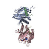
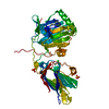
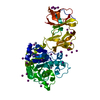
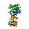
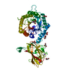
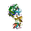
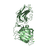
A: Ras-related protein Rab-9A B: Ras-related protein Rab-9A C: Isoform 2 of Nuclear distribution protein nudE homolog 1 D: Isoform 2 of Nuclear distribution protein nudE homolog 1 hetero molecules
In the structure databanks used in Yorodumi, some data are registered as the other names, "COVID-19 virus" and "2019-nCoV". Here are the details of the virus and the list of structure data.
Jan 31, 2019. EMDB accession codes are about to change! (news from PDBe EMDB page)
EMDB accession codes are about to change! (news from PDBe EMDB page)
The allocation of 4 digits for EMDB accession codes will soon come to an end. Whilst these codes will remain in use, new EMDB accession codes will include an additional digit and will expand incrementally as the available range of codes is exhausted. The current 4-digit format prefixed with “EMD-” (i.e. EMD-XXXX) will advance to a 5-digit format (i.e. EMD-XXXXX), and so on. It is currently estimated that the 4-digit codes will be depleted around Spring 2019, at which point the 5-digit format will come into force.
The EM Navigator/Yorodumi systems omit the EMD- prefix.
Related info.:Q: What is EMD? / ID/Accession-code notation in Yorodumi/EM Navigator
Yorodumi is a browser for structure data from EMDB, PDB, SASBDB, etc.
This page is also the successor to EM Navigator detail page, and also detail information page/front-end page for Omokage search.
The word "yorodu" (or yorozu) is an old Japanese word meaning "ten thousand". "mi" (miru) is to see.
Related info.:EMDB / PDB / SASBDB / Comparison of 3 databanks / Yorodumi Search / Aug 31, 2016. New EM Navigator & Yorodumi / Yorodumi Papers / Jmol/JSmol / Function and homology information / Changes in new EM Navigator and Yorodumi
 Movie
Movie Controller


 Open data
 Basic information
 Components
 Keywords
 Function and homology information
 Homo sapiens (human)
 X-RAY DIFFRACTION /
  Authors
 China, 1items
  Citation
 Structure visualization
 Downloads & links
 Other downloads
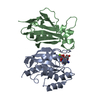


 PDBj
PDBj


 Assembly
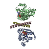


 Mass: 24.305 Da / Num. of mol.: 2 / Source method: obtained synthetically / Formula: Mg / Feature type: SUBJECT OF INVESTIGATION
 Mass: 523.180 Da / Num. of mol.: 2 / Source method: obtained synthetically / Formula: C10H16N5O14P3 / Feature type: SUBJECT OF INVESTIGATION / Comment: GTP, energy-carrying molecule*YM
 Mass: 18.015 Da / Num. of mol.: 153 / Source method: isolated from a natural source / Formula: H2O
 Sample preparation
 Processing