 Movie
Movie Controller


+ Open data

- Basic information
| Entry | Database: PDB / ID: 7e1g | |||||||||
|---|---|---|---|---|---|---|---|---|---|---|
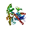
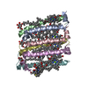
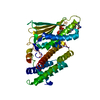
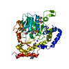
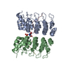
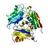
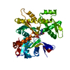
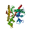
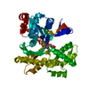
| Title | Structure of MreB3 from Spiroplasma eriocheiris | |||||||||
 Components Components | Cell shape-determining protein MreB | |||||||||
 Keywords Keywords | STRUCTURAL PROTEIN / Cytoskeleton / actin homolog | |||||||||
| Function / homology |  Function and homology information Function and homology informationcell morphogenesis / regulation of cell shape / ATP binding / metal ion binding / cytoplasm Similarity search - Function | |||||||||
| Biological species |  Spiroplasma eriocheiris (bacteria) Spiroplasma eriocheiris (bacteria) | |||||||||
| Method |  X-RAY DIFFRACTION / SYNCHROTRON / MOLECULAR REPLACEMENT / Resolution: 1.75 Å X-RAY DIFFRACTION / SYNCHROTRON / MOLECULAR REPLACEMENT / Resolution: 1.75 Å | |||||||||
 Authors Authors | Takahashi, D. / Miyata, M. / Imada, K. | |||||||||
| Funding support |  Japan, 2items Japan, 2items
| |||||||||
 Citation Citation | Journal: Open Biology / Year: 2022 Title: ATP-dependent polymerization dynamics of bacterial actin proteins involved in Spiroplasma swimming. Authors: Takahashi, D. / Fujiwara, I. / Sasajima, Y. / Narita, A. / Imada, K. / Miyata, M. | |||||||||
| History |
|
- Structure visualization
| Structure viewer | Molecule: MolmilJmol/JSmol |
|---|
- Downloads & links
-Download
| PDBx/mmCIF format | 7e1g.cif.gz | 168 KB | Display | PDBx/mmCIF format |
|---|---|---|---|---|
| PDB format | pdb7e1g.ent.gz | 129.3 KB | Display | PDB format |
| PDBx/mmJSON format | 7e1g.json.gz | Tree view | PDBx/mmJSON format | |
| Others |  Other downloads Other downloads |
-Validation report
| Summary document | 7e1g_validation.pdf.gz | 1.8 MB | Display | wwPDB validaton report |
|---|---|---|---|---|
| Full document | 7e1g_full_validation.pdf.gz | 1.8 MB | Display | |
| Data in XML | 7e1g_validation.xml.gz | 35.2 KB | Display | |
| Data in CIF | 7e1g_validation.cif.gz | 52.5 KB | Display | |
| Arichive directory | https://data.pdbj.org/pub/pdb/validation_reports/e1/7e1gftp://data.pdbj.org/pub/pdb/validation_reports/e1/7e1g | HTTPS FTP |
-Related structure data
| Related structure data | 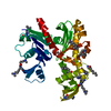 7e1cSC S: Starting model for refinement C: citing same article ( |
|---|---|
| Similar structure data |
-Links
 PDBj
PDBj


- Assembly
| Deposited unit | 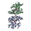
| ||||||||||
|---|---|---|---|---|---|---|---|---|---|---|---|
| 1 | 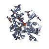
| ||||||||||
| 2 | 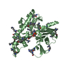
| ||||||||||
| Unit cell |
|
-Components
| #1: Protein | Mass: 39168.836 Da / Num. of mol.: 2 Source method: isolated from a genetically manipulated source Source: (gene. exp.) Spiroplasma eriocheiris (bacteria) / Gene: mreB3, mreB, SERIO_v1c12260 / Plasmid: pET15b / Production host: #2: Chemical |   Mass: 24.305 Da / Num. of mol.: 2 / Source method: obtained synthetically / Formula: Mg / Feature type: SUBJECT OF INVESTIGATION Mass: 24.305 Da / Num. of mol.: 2 / Source method: obtained synthetically / Formula: Mg / Feature type: SUBJECT OF INVESTIGATION#3: Chemical |   Mass: 506.196 Da / Num. of mol.: 2 / Source method: obtained synthetically / Formula: C10H17N6O12P3 / Feature type: SUBJECT OF INVESTIGATION / Comment: AMP-PNP, energy-carrying molecule analogue*YM Mass: 506.196 Da / Num. of mol.: 2 / Source method: obtained synthetically / Formula: C10H17N6O12P3 / Feature type: SUBJECT OF INVESTIGATION / Comment: AMP-PNP, energy-carrying molecule analogue*YM#4: Water | ChemComp-HOH / |  Mass: 18.015 Da / Num. of mol.: 763 / Source method: isolated from a natural source / Formula: H2O Mass: 18.015 Da / Num. of mol.: 763 / Source method: isolated from a natural source / Formula: H2OHas ligand of interest | Y | |
|---|
-Experimental details
-Experiment
| Experiment | Method: X-RAY DIFFRACTION / Number of used crystals: 1 |
|---|
- Sample preparation
| Crystal | Density Matthews: 2.18 Å3/Da / Density % sol: 43.5 % |
|---|---|
| Crystal grow | Temperature: 293 K / Method: vapor diffusion, sitting drop / pH: 5.8 Details: 30% (w/v) PEG 4000, 0.1 M Acetate-NaOH pH 4.6, 200 mM NH4Ac, 5 mM AMPPNP, 5mM MgCl2 |
-Data collection
| Diffraction | Mean temperature: 100 K / Serial crystal experiment: N |
|---|---|
| Diffraction source | Source: SYNCHROTRON / Site: SPring-8 / Beamline: BL41XU / Wavelength: 1 Å |
| Detector | Type: DECTRIS EIGER X 16M / Detector: PIXEL / Date: Nov 29, 2019 |
| Radiation | Monochromator: Double-crystal monochromator / Protocol: SINGLE WAVELENGTH / Monochromatic (M) / Laue (L): M / Scattering type: x-ray |
| Radiation wavelength | Wavelength: 1 Å / Relative weight: 1 |
| Reflection | Resolution: 1.75→56.3 Å / Num. obs: 66116 / % possible obs: 97.2 % / Redundancy: 3.3 % / Biso Wilson estimate: 18.86 Å2 / CC1/2: 0.993 / Rmerge(I) obs: 0.101 / Rpim(I) all: 0.065 / Net I/σ(I): 6.8 |
| Reflection shell | Resolution: 1.75→1.78 Å / Rmerge(I) obs: 0.57 / Mean I/σ(I) obs: 1.8 / Num. unique obs: 3545 / CC1/2: 0.658 / Rpim(I) all: 0.372 |
- Processing
| Software |
| |||||||||||||||||||||||||||||||||||||||||||||||||||||||||||||||||||||||||||||||||||||||||||||||||||||||||
|---|---|---|---|---|---|---|---|---|---|---|---|---|---|---|---|---|---|---|---|---|---|---|---|---|---|---|---|---|---|---|---|---|---|---|---|---|---|---|---|---|---|---|---|---|---|---|---|---|---|---|---|---|---|---|---|---|---|---|---|---|---|---|---|---|---|---|---|---|---|---|---|---|---|---|---|---|---|---|---|---|---|---|---|---|---|---|---|---|---|---|---|---|---|---|---|---|---|---|---|---|---|---|---|---|---|---|
| Refinement | Method to determine structure: MOLECULAR REPLACEMENT Starting model: 7E1C Resolution: 1.75→50.987 Å / SU ML: 0.18 / Cross valid method: FREE R-VALUE / σ(F): 1.35 / Phase error: 21.36 / Stereochemistry target values: ML
| |||||||||||||||||||||||||||||||||||||||||||||||||||||||||||||||||||||||||||||||||||||||||||||||||||||||||
| Solvent computation | Shrinkage radii: 0.9 Å / VDW probe radii: 1.11 Å / Solvent model: FLAT BULK SOLVENT MODEL | |||||||||||||||||||||||||||||||||||||||||||||||||||||||||||||||||||||||||||||||||||||||||||||||||||||||||
| Refinement step | Cycle: LAST / Resolution: 1.75→50.987 Å
| |||||||||||||||||||||||||||||||||||||||||||||||||||||||||||||||||||||||||||||||||||||||||||||||||||||||||
| Refine LS restraints |
| |||||||||||||||||||||||||||||||||||||||||||||||||||||||||||||||||||||||||||||||||||||||||||||||||||||||||
| LS refinement shell |
|
