 Movie
Movie Controller

[English] 日本語
 Yorodumi
- PDB-5j6a: Crystal structure of pyruvate dehydrogenase kinase isoform 2 in c... -
+ Open data

- Basic information
| Entry | Database: PDB / ID: 5j6a | |||||||||
|---|---|---|---|---|---|---|---|---|---|---|
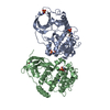
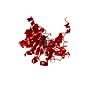
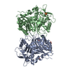
| Title | Crystal structure of pyruvate dehydrogenase kinase isoform 2 in complex with inhibitor PS46 | |||||||||
 Components Components | [Pyruvate dehydrogenase (acetyl-transferring)] kinase isozyme 2, mitochondrial | |||||||||
 Keywords Keywords | Transferase/Transferase Inhibitor / GHKL protein kinase / pyruvate dehydrogenase complex / Mitochondrial protein kinases / Impaired glucose oxidation / Hepatic steatosis / Type 2 diabetes / Cancer / Bergerat nucleotide-binding fold / protein kinase / Transferase-Transferase Inhibitor complex | |||||||||
| Function / homology |  Function and homology information Function and homology information[pyruvate dehydrogenase (acetyl-transferring)] kinase / pyruvate dehydrogenase (acetyl-transferring) kinase activity / regulation of pyruvate decarboxylation to acetyl-CoA / Regulation of pyruvate dehydrogenase (PDH) complex / regulation of ketone metabolic process / pyruvate dehydrogenase complex / regulation of pH / cellular response to nutrient / Signaling by Retinoic Acid / regulation of gluconeogenesis ...[pyruvate dehydrogenase (acetyl-transferring)] kinase / pyruvate dehydrogenase (acetyl-transferring) kinase activity / regulation of pyruvate decarboxylation to acetyl-CoA / Regulation of pyruvate dehydrogenase (PDH) complex / regulation of ketone metabolic process / pyruvate dehydrogenase complex / regulation of pH / cellular response to nutrient / Signaling by Retinoic Acid / regulation of gluconeogenesis / regulation of glucose metabolic process / intrinsic apoptotic signaling pathway by p53 class mediator / regulation of calcium-mediated signaling / cellular response to reactive oxygen species / glucose metabolic process / insulin receptor signaling pathway / glucose homeostasis / protein kinase activity / mitochondrial matrix / protein homodimerization activity / mitochondrion / nucleoplasm / ATP binding / cytosol Similarity search - Function | |||||||||
| Biological species |  Homo sapiens (human) Homo sapiens (human) | |||||||||
| Method |  X-RAY DIFFRACTION / SYNCHROTRON / MOLECULAR REPLACEMENT / Resolution: 2.045 Å X-RAY DIFFRACTION / SYNCHROTRON / MOLECULAR REPLACEMENT / Resolution: 2.045 Å | |||||||||
 Authors Authors | Gui, W.J. / Tso, S.C. / Chuang, J.L. / Wu, C.Y. / Qi, X. / Wynn, R.M. / Chuang, D.T. | |||||||||
| Funding support |  United States, 2items United States, 2items
| |||||||||
 Citation Citation | Journal: J. Med. Chem. / Year: 2017 Title: Development of Dihydroxyphenyl Sulfonylisoindoline Derivatives as Liver-Targeting Pyruvate Dehydrogenase Kinase Inhibitors. Authors: Tso, S.C. / Lou, M. / Wu, C.Y. / Gui, W.J. / Chuang, J.L. / Morlock, L.K. / Williams, N.S. / Wynn, R.M. / Qi, X. / Chuang, D.T. #1: Journal: To Be PublishedTitle: Development of Dihydroxyphenyl Sulfonylisoindoline-based Pyruvate Dehydrogenase Kinase Inhibitor Targeting Liver of Diet-induced Obese Mice Authors: Tso, S.C. / Qi, X. / Wu, C.Y. / Gui, W.J. / Chuang, J.L. / Morlock, L.K. / Williams, N.S. / Wynn, R.M. / Chuang, D.T. | |||||||||
| History |
|
- Structure visualization
| Structure viewer | Molecule: MolmilJmol/JSmol |
|---|
- Downloads & links
-Download
| PDBx/mmCIF format | 5j6a.cif.gz | 88.1 KB | Display | PDBx/mmCIF format |
|---|---|---|---|---|
| PDB format | pdb5j6a.ent.gz | 63.9 KB | Display | PDB format |
| PDBx/mmJSON format | 5j6a.json.gz | Tree view | PDBx/mmJSON format | |
| Others |  Other downloads Other downloads |
-Validation report
| Arichive directory | https://data.pdbj.org/pub/pdb/validation_reports/j6/5j6aftp://data.pdbj.org/pub/pdb/validation_reports/j6/5j6a | HTTPS FTP |
|---|
-Related structure data
| Related structure data |  5j71C  4mpnS S: Starting model for refinement C: citing same article ( |
|---|---|
| Similar structure data |


-Links
 PDBj
PDBj


- Assembly
| Deposited unit | 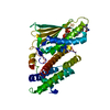
| ||||||||
|---|---|---|---|---|---|---|---|---|---|
| 1 | 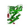
| ||||||||
| Unit cell |
|
-Components
| #1: Protein | [ Mass: 45233.402 Da / Num. of mol.: 1 Source method: isolated from a genetically manipulated source Source: (gene. exp.) Homo sapiens (human) / Gene: PDK2, PDHK2 / Production host:  References: UniProt: Q15119, [pyruvate dehydrogenase (acetyl-transferring)] kinase |
|---|---|
| #2: Chemical | ChemComp-P46 / (  Mass: 501.555 Da / Num. of mol.: 1 / Source method: obtained synthetically / Formula: C23H27N5O6S Mass: 501.555 Da / Num. of mol.: 1 / Source method: obtained synthetically / Formula: C23H27N5O6S |
| #3: Water | ChemComp-HOH /  Mass: 18.015 Da / Num. of mol.: 109 / Source method: isolated from a natural source / Formula: H2O Mass: 18.015 Da / Num. of mol.: 109 / Source method: isolated from a natural source / Formula: H2O |
-Experimental details
-Experiment
| Experiment | Method: X-RAY DIFFRACTION / Number of used crystals: 1 |
|---|
- Sample preparation
| Crystal | Density Matthews: 3.22 Å3/Da / Density % sol: 61.84 % |
|---|---|
| Crystal grow | Temperature: 293 K / Method: vapor diffusion, hanging drop / pH: 5.6 Details: 100 mM sodium acetate pH 5.6 9% Isopropanol 100 mM megnesium chloride 50 mM Ammonium Sulfate |
-Data collection
| Diffraction | Mean temperature: 100 K |
|---|---|
| Diffraction source | Source: SYNCHROTRON / Site: APS / Beamline: 19-ID / Wavelength: 0.97918 Å |
| Detector | Type: ADSC QUANTUM 315r / Detector: CCD / Date: Mar 18, 2015 |
| Radiation | Protocol: SINGLE WAVELENGTH / Monochromatic (M) / Laue (L): M / Scattering type: x-ray |
| Radiation wavelength | Wavelength: 0.97918 Å / Relative weight: 1 |
| Reflection | Resolution: 2.045→50 Å / Num. obs: 36197 / % possible obs: 99.5 % / Redundancy: 7.3 % / CC1/2: 0.741 / Rmerge(I) obs: 0.068 / Net I/σ(I): 24.7 |
| Reflection shell | Resolution: 2.05→2.09 Å / Redundancy: 7.1 % / Mean I/σ(I) obs: 2.23 / % possible all: 99.2 |
- Processing
| Software |
| |||||||||||||||||||||||||||||||||||||||||||||||||||||||||||||||||||||||||||||||||||||||||||||||||||||||||||||||||||||||||||||||||||||||||||||||||||||||||||||||||||||||||||||||
|---|---|---|---|---|---|---|---|---|---|---|---|---|---|---|---|---|---|---|---|---|---|---|---|---|---|---|---|---|---|---|---|---|---|---|---|---|---|---|---|---|---|---|---|---|---|---|---|---|---|---|---|---|---|---|---|---|---|---|---|---|---|---|---|---|---|---|---|---|---|---|---|---|---|---|---|---|---|---|---|---|---|---|---|---|---|---|---|---|---|---|---|---|---|---|---|---|---|---|---|---|---|---|---|---|---|---|---|---|---|---|---|---|---|---|---|---|---|---|---|---|---|---|---|---|---|---|---|---|---|---|---|---|---|---|---|---|---|---|---|---|---|---|---|---|---|---|---|---|---|---|---|---|---|---|---|---|---|---|---|---|---|---|---|---|---|---|---|---|---|---|---|---|---|---|---|---|
| Refinement | Method to determine structure: MOLECULAR REPLACEMENT Starting model: 4MPN Resolution: 2.045→31.513 Å / SU ML: 0.22 / Cross valid method: FREE R-VALUE / σ(F): 1.35 / Phase error: 24.58 / Stereochemistry target values: ML
| |||||||||||||||||||||||||||||||||||||||||||||||||||||||||||||||||||||||||||||||||||||||||||||||||||||||||||||||||||||||||||||||||||||||||||||||||||||||||||||||||||||||||||||||
| Solvent computation | Shrinkage radii: 0.9 Å / VDW probe radii: 1.11 Å / Solvent model: FLAT BULK SOLVENT MODEL | |||||||||||||||||||||||||||||||||||||||||||||||||||||||||||||||||||||||||||||||||||||||||||||||||||||||||||||||||||||||||||||||||||||||||||||||||||||||||||||||||||||||||||||||
| Refinement step | Cycle: LAST / Resolution: 2.045→31.513 Å
| |||||||||||||||||||||||||||||||||||||||||||||||||||||||||||||||||||||||||||||||||||||||||||||||||||||||||||||||||||||||||||||||||||||||||||||||||||||||||||||||||||||||||||||||
| Refine LS restraints |
| |||||||||||||||||||||||||||||||||||||||||||||||||||||||||||||||||||||||||||||||||||||||||||||||||||||||||||||||||||||||||||||||||||||||||||||||||||||||||||||||||||||||||||||||
| LS refinement shell |
|
